Research article

A Feasible Role of Neuropilin Signaling in Pharyngeal Pouch Formation in Zebrafish

Chong Pyo Choe1,2,https://orcid.org/0000-0003-0254-9438
Author Information & Copyright
1Division of Life Science, Gyeongsang National University, Jinju 52828, Korea
2Plant Molecular Biology and Biotechnology Research Center, Gyeongsang National University, Jinju 52828, Korea
Corresponding author Chong Pyo Choe, Division of Life Science, Gyeongsang National University, Jinju 52828, Korea Tel: +82-55-772-1367, Fax: +82-55-772-1349, E-mail: cpchoe@gnu.ac.kr

© Copyright 2023 The Korean Society of Developmental Biology. This is an Open-Access article distributed under the terms of the Creative Commons Attribution Non-Commercial License (http://creative-commons.org/licenses/by-nc/3.0/) which permits unrestricted non-commercial use, distribution, and reproduction in any medium, provided the original work is properly cited.

Received: Jul 21, 2023 ; Revised: Aug 22, 2023 ; Accepted: Sep 14, 2023

Published Online: Sep 30, 2023

Abstract

Pharyngeal pouches are an important epithelial structure controlling facial skeletal development in vertebrates. A series of pouches arise sequentially in the pharyngeal endoderm through collective cell migration followed by rearrangement of pouch-forming cells. While crucial transcription factors and signaling molecules have been identified in pouch formation, a role for Neuropilins (Nrps) in pouch development has not yet been analyzed in any vertebrates. Nrps are cell surface receptors essential for angiogenesis and axon guidance. In all vertebrates, the two Nrp family members, Nrp1 and Nrp2, are conserved in the genome, with two paralogs for Nrp1 (Nrp1a and Nrp1b) and Nrp2 (Nrp2a and Nrp2b) being identified in zebrafish. Here, I report a potential requirement of Nrp signaling in pouch development in zebrafish. nrp1a and nrp2b were expressed in the developing pouches, with sema3d, a ligand for Nrps, being expressed in the pouches. Knocking down Nrps signaling in the pharyngeal endoderm led to severe defects in pouches and facial cartilages. In addition, blocking Mitogen-activated protein kinase (MAPK) activities, a downstream effector of Nrp signaling, in the pharyngeal endoderm caused similar defects in pouches and facial skeleton to those by knocking down Nrps signaling. My results suggest that Nrp signaling acts for pouch formation through MAPK.

Keywords: Neuropilin; Pharyngeal pouch; Facial cartilages; Craniofacial development; Zebrafish

INTRODUCTION

The pharyngeal pouches are a transient epithelial structure essential for craniofacial development in vertebrates. They arise in the pharyngeal endoderm as a series of branches, which segment the pharyngeal arches and provide survival signals, such as sonic Hedgehog, to the cranial neural crest cells (cNCCs) in the arches. The segmented cNCCs then differentiate into the facial skeletal elements, including facial cartilages (Mork & Crump, 2015). The pouches, per se, further differentiate into endocrine glands in the head and neck, including the thymus and parathyroid (Grevellec & Tucker, 2010). Consistent with essential roles for the pouches in craniofacial development, defects in pouches led to severe loss or malformation of facial cartilages as well as hypoplasia of the thymus and parathyroid in mice and zebrafish (Piotrowski & Nusslein-Volhard, 2000; Dickmeis et al., 2001; Kikuchi et al., 2001; Grevellec & Tucker, 2010). In humans, abnormal pouches are an etiology of DiGeorge syndrome, a congenital craniofacial disability (Driscoll et al., 1992; Piotrowski & Nusslein-Volhard, 2000; Lindsay et al., 2001; Tran et al., 2011). Given the importance of pouches in craniofacial development, the genetic and cellular mechanisms controlling pouch formation are less understood in vertebrates. Genetically, Tbx1, Pax1, and Foxi1 transcription factors and Fgf, Wnt, Ephrin, and VEGF-C signaling molecules are essential for pouch formation (Lindsay et al., 2001; Nissen et al., 2003; Piotrowski et al., 2003; Crump et al., 2004; Ober et al., 2004; Tran et al., 2011; Choe & Crump, 2014; Okada et al., 2016; Jin et al., 2018; Liu et al., 2020). In zebrafish, Tbx1 initiates pouch formation by promoting a collective migration of pouch-forming cells through Wnt11r and Fgf8a (Choe et al., 2013; Choe & Crump, 2014). Migrating pouch-forming cells are then rearranged into a bi-layered structure by Wnt4 and Ephrin B2 and B3 ligands to mature pouches, with wnt4a expression being regulated by Foxi1 in zebrafish (Choe et al., 2013; Choe & Crump, 2015; Jin et al., 2018). In medaka and zebrafish, Pax1 regulates the expression of tbx1 and fgf3 in the pharyngeal endoderm to control pouch formation (Okada et al., 2016; Liu et al., 2020). Besides these transcription factors and signaling molecules, barely anything has been identified in pouch formation in vertebrates. Here, I tested the potential role of Neuropilins (Nrps) in developing pouches in zebrafish.

The Nrps are a family of cell surface receptors with many critical biological functions in the cardiovascular and neuronal systems (Guo & Vander Kooi, 2015). In vertebrates, two Npr family members, Nrp1 and Nrp2, are conserved and transduce VEGF and Semaphorin signals in the cardiovascular and neuronal systems (Guo & Vander Kooi, 2015). In angiogenesis, Nrps form a ligand/receptor complex, including VEGF and VEGFR (Guo & Vander Kooi, 2015). Nrp1 interacts with VEGF-A and VEGFR2 in mice and controls endothelial cell migration (Soker et al., 1998). In particular, it functions in endothelial tip cells as a downstream effector of a Notch signaling pathway (Fantin et al., 2013). The knockout of the Nrp1 gene in mice consistently led to embryonic lethality due to severe defects in endothelial cell migration (Jones et al., 2008). In zebrafish, knockdown of nrp1 has shown similar defects in angiogenesis (Lee et al., 2002). Nrp2 acts with VEGF-C and VEGFR2/3 in mice and is important for lymph angiogenesis (Yuan et al., 2002; Kärpänen et al., 2006). Npr2 functions in lymphatic tip cells and modulates tip cell extension at the cellular level (Caunt et al., 2008; Xu et al., 2010). Although a functional validation has remained, zebrafish Nrp2 also has been implicated in lymph angiogenesis as it is expressed in lymphatic vessels during embryogenesis (Yaniv et al., 2006). To transduce Nrp signals in endothelial cells, Mitogen-activated protein kinase (MAPK) has been implicated as a critical downstream effector of Nrp1 and Nrp2 (Wey et al., 2005; Bechara et al., 2008; Fung et al., 2016). Although the roles of Npr1 and Nrp2 are distinct, it has been implicated that they can compensate for each other in angiogenesis as the double knockout mouse displayed more severe defects in angiogenesis (Takashima et al., 2002). Besides VEGFs, Nrps are receptors for Semaphorin3 (Sema3) ligands. They act with the Plexin receptor to bind Sema3 and control axon guidance in the neuronal system (Parker et al., 2012). Sema3 ligands have been demonstrated as endogenous inhibitors of angiogenesis and lymphogenesis through Nrps (Mumblat et al., 2015; Nakayama et al., 2015; Yang et al., 2015). Indeed, Nrps integrate competitive VEGF and Sema3 signals to control endothelial cell migration (Serini et al., 2003; Tamagnone & Mazzone, 2011).

At the cellular level, the directed collective migration of endodermal cells initiates pouch formation, which is similar to the collective migration of endothelial cells in angiogenesis (Costa et al., 2016). Although VEGFRs have not been identified yet, the Vegf-C ligand is required for pouch formation in zebrafish (Ober et al., 2004). Furthermore, I found that Nrps and a Sema3 ligand were expressed in the pharyngeal endoderm during pouch formation in zebrafish. Based on this observation, I tested the potential role of Nrp signaling in pouch morphogenesis. Blocking the Nrp signal, specifically in the pharyngeal endoderm during pouch formation, led to severe defects in pouches and facial cartilages whose formation depended on appropriate pouch development. Knockdown of MAPK activities in the endoderm demonstrated defects in pouches and facial skeletons. This preliminary functional analysis suggests that Nrps are able to function in the pharyngeal endoderm to form pouches through MAPK.

MATERIALS AND METHODS

1. Zebrafish lines

Zebrafish were handled according to the Animal Protection Act (2017) in Korea. All zebrafish work was approved by Gyeongsang National University Institutional Animal Care and Use Committee. Published lines include Tg(~3.4her5:EGFP) (Tallafuss & Bally-Cuif, 2003) and Tg(nkx2.3:Gal4VP16) (Choe et al., 2013). Tg(UAS:DN-Nrp1a), Tg(UAS:Sema3d), and Tg(UAS:DN-MAPK3) transgenic constructs were generated using the Gateway (Invitrogen, Carlsbad, CA, USA) Tol2kit (Kwan et al., 2007). For pME-DN-Nrp1a, a dominant-negative form of Nrp1a that is missing its cytoplasmic domain was produced by PCR using primers DN-Nrp1a-B1F2 and DN-Nrp1a-B2R from multi-stage zebrafish cDNA (Renzi et al., 1999). For pME-Sema3d, the coding sequence of sema3d was amplified using primers Sema3d-B1F2 and Sema3d-B2R from multi-stage zebrafish cDNA. For pME-DN-MAPK3, a dominant-negative form of MAPK3 was produced by fusion PCR with primers designed to produce a lysine to arginine substitution at lysine 85 (Izumi et al., 2001). 30 ng of transgenic constructs and 35 ng of DNA construct bearing Tol2 transposase gene were injected into one-cell stage wild-type Tübingen embryos. Three independent transgenic lines for Tg(UAS:DN-Nrp1a:pA), two for Tg(UAS:Sema3d:pA), and three Tg(UAS:DN-MAPK3:pA) were established based on α-crystallin:Cerulean eye fluorescence. Tg(UAS:DN-Nrp1a:pA)GNU125, Tg(UAS:Sema3d:pA)GNU141, and Tg(UAS:DN-MAPK3:pA)GNU152 were used for this study. Primers are listed in Table 1.

Table 1. List of primers used to create transgenic lines
Primer Sequences (5’ to 3’)
DN-Nrp1a_F ATG CAT TGT GGA TTA GTG
DN-Nrp1a_MR ACT CAT GGC GAT GAT AGT TGT AGG CAC TTC CAT TTC
DN-Nrp1a_MF GAA ATG GAA GTG CCT ACA ACT ATC ATC GCC ATG AGT
DN-Nrp1a_R TCA CGC TTC CGA GTA CGA
DN-Nrp1a_B1F2 GGGGACAAGTTTGTACAAAAAAGCAGGCT ATG CAT TGT GGA TTA GTG TTG
DN-Nrp1a_B2R GGGGACCACTTTGTACAAGAAAGCTGGGT TCA CGC TTC CGA GTA CGA
Sema3d_B1F2 GGGGACAAGTTTGTACAAAAAAGCAGGCT ATG AAG ACT GCA GGG GAG
Sema3d_B2R GGGGACCACTTTGTACAAGAAAGCTGGGT CTA GTG GTG GCG TCG GTT TCT
DN-MAPK3_F ATG GCG GAA TCG GGC AGT
DN-MAPK3_MR CAA ATG GAC TTA TCT TCT TAA TGG CCA CTC GGA TCT
DN-MAPK3_MF AGA TCC GAG TGG CCA TTA AGA AGA TAA GTC CAT TTG
DN-MAPK3_F TCA GGA GCC CTG GTA ATT
DN-MAPK3_B1F2 GGGGACAAGTTTGTACAAAAAAGCAGGCT ATG GCG GAA TCG GGC AGT
DN-MAPK3_B2R GGGGACCACTTTGTACAAGAAAGCTGGGT TCA GGA GCC CTG GTA ATT

* Att sites are underlined.

* MAPK, mitogen-activated protein kinase.

Download Excel Table
2. Staining

Immunohistochemistry for Zn8 (Zebrafish International Resource Center, 1:400), fluorescent in situ hybridization in conjunction with GFP immunohistochemistry (Torrey Pines Biolabs, Secaucus, NJ, USA; 1:1,000), and Alcian blue staining were carried out as described previously (Crump et al., 2004; Zuniga et al., 2011). Partial cDNA fragments of nrp1a, nrp2b, and sema3d were amplified from mixed-stage zebrafish cDNA. The PCR fragments were cloned into the pGEM®-T easy vector (Promega, Madison, WI, USA) and were confirmed by DNA sequencing. Digoxigenin (DIG)-labeled antisense riboprobes were transcribed with T7/SP6 RNA polymerase (Roche Life Sciences, Basel, Switzerland) from linearized plasmids. Primers used for riboprobes are listed in Table 2.

Table 2. List of primers used to create in situ probes
Gene Forward primer (5’ to 3’) Reverse primer (5’ to 3’)
nrp1a CAG TCA CAA CCG TAT GCA CGA AGG ATC ATT TGC CCA
nrp1b TAT CTA CCC TGA GCG TGG TCC GTC TCC ATC TCA TCC
nrp2a GAG CTC CAG GGA ATG TTG GTT GTG TTG GTT TGC TCG
nrp2b AGA ACT TTA CGG CTG TCA TGG TGG TAG ATT CTG GCA
sema3d TTG GGC ACA GAT ATG GGA CAC GCT CGT CTG GTT TAA
Download Excel Table
3. Imaging

An Olympus FLUOVIEW FV3000 confocal microscope using FV31S-SW software (Olympus, Tokyo, Japan) captured fluorescent images for immunohistochemistry and in situ hybridization. Approximately 100 μm Z-stacks at 3.5-μm intervals were taken with an Olympus UPLXAPO 20X objective lens, then were projected into a single image. An Olympus BX50 upright microscope using mosaic V2.1 software (Tucsen Photonics, Fuzhou, China) imaged flat-mounted facial cartilages dissected manually from Alcian Blue staining larvae. Any adjustments were applied to all panels using Adobe Photoshop (Adobe Systems, San Jose, CA, USA).

4. Statistics

Pouches and ceratobranchial (Cb) cartilages were scored to quantify defects in pouches and Cb cartilages. The normal pouch was scored as 1, abnormal and hypoplasic as 0.5, and missing as 0. Similarly, normal Cb was scored as 1, hypoplasic or fused Cb as 0.5, and missing Cb as 0. The multiple comparison test of Tukey–Kramer was employed to quantify defects.

RESULTS

1. Zebrafish nrp1a, nrp2b, and sema3d are expressed in pharyngeal endoderm forming pharyngeal pouches

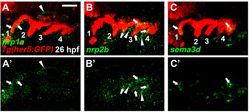
There are four nrp genes, nrp1a, nrp1b, nrp2a, and nrp2b, in zebrafish. Previously, it has been reported that all four nrp genes are expressed in the brain at 24 to 72 hours-post-fertilization (hpf), with nrp1a and nrp2b being expressed specifically in mandibular cartilages at 36 to 72 hpf (Bovenkamp et al., 2004). However, the expression of nrp genes in the pharyngeal regions of the embryonic head has yet to be analyzed. To investigate a potential role of Nrp signaling in craniofacial development, I first examined the expression of nrp genes in the pharyngeal endoderm at 26 hpf, in which a series of pouches arises (Choe et al., 2013). To do so, I carried out in situ hybridization for nrp genes in wild-type embryos harboring Tg(her5:GFP) transgene, a reporter of the pharyngeal endoderm and pouches (Tallafuss & Bally-Cuif, 2003). When the posterior pouches formed at 26 hpf, nrp1a was expressed in the first pouch, with its expression not seen in other pouches (arrows in Fig. 1A). In addition, its expression was seen in the brain (arrowhead in Fig. 1A) and heart (asterisk in Fig. 1A). Transcripts for nrp2b were observed in the posterior pouches, including the second to fourth pouches at 26 hpf, whereas they were not seen in the first pouch (arrows in Fig. 1B). Besides, nrp2b was also expressed in the adjacent mesoderm to the pouches (arrowheads in Fig. 1B). Apparently, nrp1b and nrp2a were not expressed in the pharyngeal regions at 26 hpf. Next, I analyzed the expression of sema3 genes, ligands for Nrp receptors, in the endoderm during pouch formation. Among sema3 genes, I found that sema3d was expressed in the matured pouches as well as in the pharyngeal endoderm forming the fifth pouch at 26 hpf (arrows in Fig. 1C). Expression of nrp1a, nrp2b, and sema3d in the pharyngeal pouches at 26 hpf suggests that they could work together to form pouches during craniofacial development.
dr-27-3-137-g1
Fig. 1. Expression of nrp1a, nrp2b, and sema3d at 26 hpf. (A–C) Fluorescent in situ hybridization (green) in conjunction with the GFP immunohistochemistry (red) in wild-type Tg(her5:GFP) animals. (A) Arrows indicate nrp1a expression in the her5-positive first (1) pouch. nrp1a expression in other pouches is not seen. Expression of nrp1a in the brain and heart is marked with an arrowhead and asterisk, respectively. (B) nrp2b expression in the her5-positive second (2) to fourth (4) pouches is marked with arrows, with its expression in the adjacent mesoderm being indicated with arrowheads. (C) sema3d expression in the her5-positive first pouch (1) and developing fourth pouch (4) is marked with arrows. (A’–C’) Green channel only. Pouches are numbered. Scale bars: 40 μm. Anterior is to the left.
Download Original Figure
2. Nrp signaling is required in the pharyngeal endoderm to develop pharyngeal pouches

I utilized a dominant-negative approach to access the function of Nrp signaling in pouch formation. To do so, I generated Tg(UAS:DN-Nrp1a) lines harboring a dominant-negative mutant allele of nrp1a. Utilizing the pharyngeal endoderm driver Tg(nkx2.3:Gal4VP16) lines that are able to express target genes in pouch-forming endoderm, including the third and fourth pouches (Choe et al., 2013), I overexpressed DN-Nrp1a specifically in the endoderm during pouch formation. Overexpression of the DN-Nrp1a in the endoderm was expected for the endodermal cells to prevent them from receiving signals mediated by Nrps, including Nrp1a. In wild types, five pouches were visualized by Zn8 immunostaining at 34 hpf (Fig. 2A and D). However, compound animals bearing Tg(nkx2.3:Gal4VP16) and Tg(UAS:DN-Nrp1a) transgenes displayed abnormal pouches at 34 hpf (Fig. 2B). Although the first pouch was normal, the remaining pouches were hypoplasic, misshaped, or missing (Fig. 2B and D). In addition, pouches 3–5 were not separated from each other (arrows in Fig. 2B). This result suggests that Nrp signals are required in pharyngeal endoderm for pouch formation. I further verified the role of Nrp signals in pouch development by analyzing Cb cartilages whose formation relied on normal pouch development (Crump et al., 2004). Five pairs of Cbs formed symmetrically in wild types at 4 dpf (dots in Fig. 3A and D). In the compound animals, however, Cbs were missing or fused, confirming defects in pouches (arrows in Fig. 3B and D). Taken together with the expression of nrp1a and nrp2b in the endoderm and pouches during pouch formation, my functional analysis suggests a critical role of Nrp signals in developing pouches and facial cartilages.

dr-27-3-137-g2
Fig. 2. Requirement of Nrp-Sema3 signaling in pharyngeal endoderm for pouch formation. (A–C) Zn8 immunohistochemistry (green) visualizes pouches at 34 hpf. (A) Zn8 staining labels five pouches in wild types. (B) In compound animals bearing Tg(nkx2.3:Gal4VP16) and Tg(UAS:DN-Nrp1a) transgenes, pouches 2, 4, and 5 are malformed, with the first (1) and third (3) pouches being fairly normal. Arrows indicate pouches 3–5 that are not separated completely. (C) Overexpression of Sema3d in nkx2.3-positive endoderm formed abnormal first (1) and fifth (5) pouches, with the fifth one not completely separated from the fourth one (arrow). Scale bars: 40 μm. Anterior is to the left. (D) Quantification of pouch defects. Number of pouches examined for each genotype: wild type (37), Tg(nkx2.3:Gal4VP16); Tg(UAS:DN-Nrp1a) (45), Tg(nkx2.3:Gal4VP16); Tg(UAS:Sema3d) (41). Data represent on a boxplot. *** p<0.001.
Download Original Figure
dr-27-3-137-g3
Fig. 3. Requirement of Nrp-Sema3 signaling in developing ceratobranchial cartilages (Cbs). (A) Dots indicate five pairs of Cbs in wild types at 4 dpf. (B) Arrows mark Cbs fused to each other, with dots marking normal Cbs in compound animals bearing Tg(nkx2.3:Gal4VP16) and Tg(UAS:DN-Nrp1a) transgenes. (C) Overexpression of Sema3d in nkx2.3-positive endoderm displays hypoplasic (red arrow) and fused (arrows) Cbs. Normal Cbs are marked with dots. Anterior is to the left. (D) Quantification of Cb defects. Number of Cbs examined for each genotype: wild type (31), Tg(nkx2.3:Gal4VP16); Tg(UAS:DN-Nrp1a) (39), Tg(nkx2.3:Gal4VP16); Tg(UAS:Sema3d) (36). Data represent on a boxplot. *** p<0.001.
Download Original Figure
3. Sema3d is able to control pouch formation

As I observed sema3d expression in developing pouches at 26 hpf, I also tested the potential role of Sema3d in pouch formation. To do so, I expressed sema3d ectopically in the pharyngeal endoderm during pouch formation by the UAS/Gal4 system. In compound transgenic animals harboring Tg(nkx2.3:Gal4VP16) and Tg(UAS:Sema3d) transgenes, pouches were missing or malformed compared to wild types (Fig. 2A, C, and D). For example, the first pouch was barely seen, and the fifth one was not completely separated from the fourth one (Fig. 2C). Consistent with defects in pouches, Cb cartilages were also fused to other Cbs or malformed in the compound animals (Fig. 3C and D). This result shows that the endodermal cells are able to respond to ectopically expressed sema3d during pouch formation, implying a potential role of Sema3d in pouch formation.

4. Development of pouches requires mitogen-activated protein kinase (MAPK) activities in the endoderm

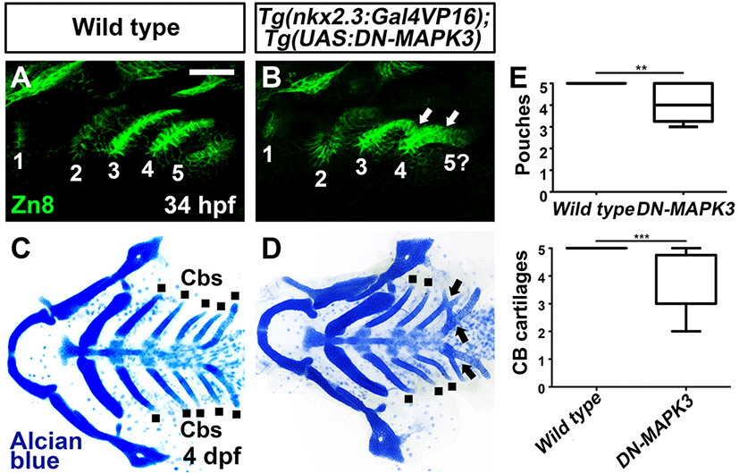
In the cellular contexts, activation of Nrp receptors promotes MAPK signaling, with Sema3 family ligands being required for MAPK activation (Wey et al., 2005; Bechara et al., 2008; Fung et al., 2016; Hai et al., 2022). Since I observed a potential requirement of the Nrp-Sema3d signal in pouch formation, I next examined whether MAPK was necessary in the endoderm for pouch development. To knock down MAPK activities in the endoderm during pouch formation, I overexpressed a dominant-negative form of MAPK3, specifically in the endoderm with the UAS/Gal4 system. Compared to wild types, compound Tg(nkx2.3:Gal4VP16) and Tg(UAS:DN-MAPK3) transgenic animals showed defects in pouches, including missing pouch and incompletely separated pouches (Fig. 4A, B, and E), and in Cbs such as fusion among Cbs (Fig. 4C–E). Interestingly, these defects were similar to those seen in transgenic animals knocking down Nrp signals in pharyngeal endoderm (Fig. 2B), implying a genetic interaction between Nrp and MAPK signals in the endoderm for pouch formation.
dr-27-3-137-g4
Fig. 4. Development of pharyngeal pouches and facial cartilages in compound animals harboring Tg(nkx2.3:Gal4VP16) and Tg(UAS:DN-MAPK3) transgenes. (A, B) Zn8 staining labels to visualize pouches. Compared to wild types, overexpression of DN-MAPK3 in nkx2.3-positive endoderm shows a missing fifth pouch (5?), with incompletely segmented pouches (arrows in B). (C, D) Ventral of dissected facial cartilages. While the wild type displays the bilateral set of five Cb cartilages (dots), overexpression of DN-MAPK3 in nkx2.3-positive endoderm shows fused Cbs (arrows). Scale bar: 40 μm. Anterior is to the left. (E) Quantification of pouch and Cb defects. Number of pouches and Cb examined for each genotype: wild type (34, 37), Tg(nkx2.3:Gal4VP16); Tg(UAS:MAPK3) (39, 32). Data represent on a boxplot. *** p<0.001. Cb, ceratobranchial cartilage; MAPK, mitogen-activated protein kinase.
Download Original Figure

DISCUSSION

In this study, I examined a potential requirement of the Sema3d-Nrp-MAPK signal in pouch formation in zebrafish. Expression of nrp1a, nrp2b, and sema3d in pharyngeal endoderm, including pouches, suggests they could be necessary for pouch formation. Indeed, knockdown of Nrp signals or ectopic expression of Sema3d, specifically in the endoderm during pouch formation, resulted in defects in pouches and facial cartilages whose formation was dependent upon normal pouch formation. Furthermore, preventing MAPK activities in the endoderm showed similar defects in pouches and facial cartilages to those knocking down Nrp signals. Thus, it is feasible that the Sema3d-Nrp-MAPK signal is a critical signaling pathway controlling pouch formation in zebrafish. However, my transgenic analysis of the signal is unable to determine the role of the Sema3d-Nrp-MAPK signal in pouch formation for the following reasons. First, the dominant negative approach could knock down activities of Nrp family proteins simultaneously, but it cannot identify nrp genes necessary for pouch formation. Thus, to determine the role of Nrp signals in pouch formation, a loss-of-function (LOF) analysis of nrp1a and nrp2b genes using LOF mutants is required. Second, similar to the first reason, the Sema3d-Nrp signal cannot be determined in pouch formation without LOF analysis of sema3d. In particular, instead of Sema3d, Vegf-C that has been implicated in pouch formation could function in pouch formation through Nrps, as Vegf can transduce signals through heterodimer for VEGFR and Nrp (Soker et al., 1998; Ober et al., 2004; Guo & Vander Kooi, 2015). However, VEGFRs required for pouch formation have yet to be identified in zebrafish. Third, although MAPKs are essential downstream effectors to transduce Nrp signals, they are involved in transducing several signals from crucial growth factors, including EGF, TGF, and VEGF, in various cellular contexts (Zhang & Liu, 2002). Thus, analyzing genetic or molecular interactions between Nrps and MAPKs in pouch formation is necessary for determining the role of the Nrp-MAPK signal in pouch formation. Although it is still preliminary to identify a new signal to control pouch formation, my study suggests a possible role of the Sema3d-Nrp-MAPK signal in pouch formation. Currently, I am developing genetic and molecular tools to test the Sema3d-Nrp-MAPK signal in pouch formation at the genetic and molecular levels.

Conflict of interests

The authors declare no potential conflict of interest.

Acknowledgements

I thank Sil Jin for technical assistance. This work was supported by a grant from Basic Science Research Program through the National Research Foundation of Korea (NRF) funded by the Ministry of Science and ICT (2022R1F1A1060199).

Authors’ contributions

The article is prepared by a single author.

Ethics approval

Animal care and experimental procedures were approved by Gyeongsang National University Institutional Animal Care and Use Committee in accordance with guidelines established by the Korea Food and Drug Administration (GNU-201012-E0074).

REFERENCES

1.

Bechara A, Nawabi H, Moret F, Yaron A, Weaver E, Bozon M, Abouzid K, Guan JL, Tessier-Lavigne M, Lemmon V, Castellani V. 2008; FAK-MAPK-dependent adhesion disassembly downstream of L1 contributes to semaphorin3A-induced collapse. EMBO J. 27:1549-1562

2.

Bovenkamp DE, Goishi K, Bahary N, Davidson AJ, Zhou Y, Becker T, Becker CG, Zon LI, Klagsbrun M. 2004; Expression and mapping of duplicate neuropilin-1 and neuropilin-2 genes in developing zebrafish. Gene Expr Patterns. 4:361-370

3.

Caunt M, Mak J, Liang WC, Stawicki S, Pan Q, Tong RK, Kowalski J, Ho C, Reslan HB, Ross J, Berry L, Kasman I, Zlot C, Cheng Z, Le Couter J, Filvaroff EH, Plowman G, Peale F, French D, Carano R, Koch AW, Wu Y, Watts RJ, Tessier-Lavigne M, Bagri A. 2008; Blocking neuropilin-2 function inhibits tumor cell metastasis. Cancer Cell. 13:331-342

4.

Choe CP, Collazo A, Trinh LA, Pan L, Moens CB, Crump JG. 2013; Wnt-dependent epithelial transitions drive pharyngeal pouch formation. Dev Cell. 24:296-309

5.

Choe CP, Crump JG. 2014; Tbx1 controls the morphogenesis of pharyngeal pouch epithelia through mesodermal Wnt11r and Fgf8a. Development. 141:3583-3593

6.

Choe CP, Crump JG. 2015; Eph-Pak2a signaling regulates branching of the pharyngeal endoderm by inhibiting late-stage epithelial dynamics. Development. 142:1089-1094

7.

Costa G, Harrington KI, Lovegrove HE, Page DJ, Chakravartula S, Bentley K, Herbert SP. 2016; Asymmetric division coordinates collective cell migration in angiogenesis. Nat Cell Biol. 18:1292-1301

8.

Crump JG, Maves L, Lawson ND, Weinstein BM, Kimmel CB. 2004; An essential role for Fgfs in endodermal pouch formation influences later craniofacial skeletal patterning. Development. 131:5703-5716

9.

Dickmeis T, Mourrain P, Saint-Etienne L, Fischer N, Aanstad P, Clark M, Strähle U, Rosa F. 2001; A crucial component of the endoderm formation pathway, CASANOVA, is encoded by a novel sox-related gene. Genes Dev. 15:1487-1492

10.

Driscoll DA, Budarf ML, Emanuel BS. 1992; A genetic etiology for DiGeorge syndrome: Consistent deletions and microdeletions of 22q11. Am J Hum Genet. 50:924-933.

11.

Fantin A, Vieira JM, Plein A, Denti L, Fruttiger M, Pollard JW, Ruhrberg C. 2013; NRP1 acts cell autonomously in endothelium to promote tip cell function during sprouting angiogenesis. Blood. 121:2352-2362

12.

Fung TM, Ng KY, Tong M, Chen JN, Chai S, Chan KT, Law S, Lee NP, Choi MY, Li B, Cheung AL, Tsao SW, Qin YR, Guan XY, Chan KW, Ma S. 2016; Neuropilin-2 promotes tumourigenicity and metastasis in oesophageal squamous cell carcinoma through ERK-MAPK-ETV4-MMP-E-cadherin deregulation. J Pathol. 239:309-319

13.

Grevellec A, Tucker AS. 2010; The pharyngeal pouches and clefts: Development, evolution, structure and derivatives. Semin Cell Dev Biol. 21:325-332

14.

Guo HF, Vander Kooi CW. 2015; Neuropilin functions as an essential cell surface receptor. J Biol Chem. 290:29120-29126

15.

Hai R, You Q, Wu F, Qiu G, Yang Q, Shu L, Xie L, Zhou X. 2022; Semaphorin 3D inhibits proliferation and migration of papillary thyroid carcinoma by regulating MAPK/ERK signaling pathway. Mol Biol Rep. 49:3793-3802

16.

Izumi Y, Kim S, Namba M, Yasumoto H, Miyazaki H, Hoshiga M, Kaneda Y, Morishita R, Zhan Y, Iwao H. 2001; Gene transfer of dominant-negative mutants of extracellular signal-regulated kinase and c-Jun NH2-terminal kinase prevents neointimal formation in balloon-injured rat artery. Circ Res. 88:1120-1126

17.

Jin S, O J, Stellabotte F, Choe CP. 2018; Foxi1 promotes late-stage pharyngeal pouch morphogenesis through ectodermal Wnt4a activation. Dev Biol. 441:12-18

18.

Jones EAV, Yuan L, Breant C, Watts RJ, Eichmann A. 2008; Separating genetic and hemodynamic defects in neuropilin 1 knockout embryos. Development. 135:2479-2488

19.

Kärpänen T, Heckman CA, Keskitalo S, Jeltsch M, Ollila H, Neufeld G, Tamagnone L, Alitalo K. 2006; Functional interaction of VEGF-C and VEGF-D with neuropilin receptors. FASEB J. 20:1462-1472

20.

Kikuchi Y, Agathon A, Alexander J, Thisse C, Waldron S, Yelon D, Thisse B, Stainier DYR. 2001; casanova Encodes a novel Sox-related protein necessary and sufficient for early endoderm formation in zebrafish. Genes Dev. 15:1493-1505

21.

Kwan KM, Fujimoto E, Grabher C, Mangum BD, Hardy ME, Campbell DS, Parant JM, Joseph Yost H, Kanki JP, Chien CB. 2007; The Tol2kit: A multisite gateway-based construction kit for Tol2 transposon transgenesis constructs. Dev Dyn. 236:3088-3099

22.

Lee P, Goishi K, Davidson AJ, Mannix R, Zon L, Klagsbrun M. 2002; Neuropilin-1 is required for vascular development and is a mediator of VEGF-dependent angiogenesis in zebrafish. Proc Natl Acad Sci USA. 99:10470-10475

23.

Lindsay EA, Vitelli F, Su H, Morishima M, Huynh T, Pramparo T, Jurecic V, Ogunrinu G, Sutherland HF, Scambler PJ, Bradley A, Baldini A. 2001; Tbx1 haploinsufficieny in the DiGeorge syndrome region causes aortic arch defects in mice. Nature. 410:97-101

24.

Liu YH, Lin TC, Hwang SPL. 2020; Zebrafish Pax1a and Pax1b are required for pharyngeal pouch morphogenesis and ceratobranchial cartilage development. Mech Dev. 161:103598

25.

Mork L, Crump JG. 2015; Chapter ten - Zebrafish craniofacial development: A window into early patterning. Curr Top Dev Biol. 115:235-269

26.

Mumblat Y, Kessler O, Ilan N, Neufeld G. 2015; Full-length semaphorin-3C is an inhibitor of tumor lymphangiogenesis and metastasis. Cancer Res. 75:2177-2186

27.

Nakayama H, Bruneau S, Kochupurakkal N, Coma S, Briscoe DM, Klagsbrun M. 2015; Regulation of mTOR signaling by semaphorin 3F-neuropilin 2 interactions in vitro and in vivo. Sci Rep. 5:11789

28.

Nissen RM, Yan J, Amsterdam A, Hopkins N, Burgess SM. 2003; Zebrafish foxi one modulates cellular responses to Fgf signaling required for the integrity of ear and jaw patterning. Development. 130:2543-2554

29.

Ober EA, Olofsson B, Mäkinen T, Jin SW, Shoji W, Koh GY, Alitalo K, Stainier DYR. 2004; Vegfc is required for vascular development and endoderm morphogenesis in zebrafish. EMBO Rep. 5:78-84

30.

Okada K, Inohaya K, Mise T, Kudo A, Takada S, Wada H. 2016; Reiterative expression of pax1 directs pharyngeal pouch segmentation in medaka. Development. 143:1800-1810

31.

Parker MW, Guo HF, Li X, Linkugel AD, Vander Kooi CW. 2012; Function of members of the neuropilin family as essential pleiotropic cell surface receptors. Biochemistry. 51:9437-9446

32.

Piotrowski T, Nüsslein-Volhard C. 2000; The endoderm plays an important role in patterning the segmented pharyngeal region in zebrafish (Danio rerio). Dev Biol. 225:339-356

33.

Piotrowski T, Ahn D, Schilling TF, Nair S, Ruvinsky I, Geisler R, Rauch GJ, Haffter P, Zon LI, Zhou Y, Foott H, Dawid IB, Ho RK. 2003; The zebrafish van gogh mutation disrupts tbx1, which is involved in the DiGeorge deletion syndrome in humans. Development. 130:5043-5052

34.

Renzi MJ, Feiner L, Koppel AM, Raper JA. 1999; A dominant negative receptor for specific secreted semaphorins is generated by deleting an extracellular domain from neuropilin-1. J Neurosci. 19:7870-7880

35.

Serini G, Valdembri D, Zanivan S, Morterra G, Burkhardt C, Caccavari F, Zammataro L, Primo L, Tamagnone L, Logan M, Tessier-Lavigne M, Taniguchi M, Püschel AW, Bussolino F. 2003; Class 3 semaphorins control vascular morphogenesis by inhibiting integrin function. Nature. 424:391-397

36.

Soker S, Takashima S, Miao HQ, Neufeld G, Klagsbrun M. 1998; Neuropilin-1 is expressed by endothelial and tumor cells as an isoform-specific receptor for vascular endothelial growth factor. Cell. 92:735-745

37.

Takashima S, Kitakaze M, Asakura M, Asanuma H, Sanada S, Tashiro F, Niwa H, Miyazaki Ji J, Hirota S, Kitamura Y, Kitsukawa T, Fujisawa H, Klagsbrun M, Hori M. 2002; Targeting of both mouse neuropilin-1 and neuropilin-2 genes severely impairs developmental yolk sac and embryonic angiogenesis. Proc Natl Acad Sci USA. 99:3657-3662

38.

Tallafuß A, Bally-Cuif L. 2003; Tracing of her5 progeny in zebrafish transgenics reveals the dynamics of midbrain-hindbrain neurogenesis and maintenance. Development. 130:4307-4323.

39.

Tamagnone L, Mazzone M. 2011; Semaphorin signals on the road of endothelial tip cells. Dev Cell. 21:189-190

40.

Tran HT, Delvaeye M, Verschuere V, Descamps E, Crabbe E, Van Hoorebeke L, McCrea P, Adriaens D, Van Roy F, Vleminckx K. 2011; ARVCF depletion cooperates with Tbx1 deficiency in the development of 22q11.2DS-like phenotypes in Xenopus. Dev Dyn. 240:2680-2687

41.

Wey JS, Gray MJ, Fan F, Belcheva A, McCarty MF, Stoeltzing O, Somcio R, Liu W, Evans DB, Klagsbrun M, Gallick GE, Ellis LM. 2005; Overexpression of neuropilin-1 promotes constitutive MAPK signalling and chemoresistance in pancreatic cancer cells. Br J Cancer. 93:233-241

42.

Xu Y, Yuan L, Mak J, Pardanaud L, Caunt M, Kasman I, Larrivée B, del Toro R, Suchting S, Medvinsky A, Silva J, Yang J, Thomas JL, Koch AW, Alitalo K, Eichmann A, Bagri A. 2010; Neuropilin-2 mediates VEGF-C-induced lymphatic sprouting together with VEGFR3. J Cell Biol. 188:115-130

43.

Yang WJ, Hu J, Uemura A, Tetzlaff F, Augustin HG, Fischer A. 2015; Semaphorin-3C signals through Neuropilin-1 and PlexinD1 receptors to inhibit pathological angiogenesis. EMBO Mol Med. 7:1267-1284

44.

Yaniv K, Isogai S, Castranova D, Dye L, Hitomi J, Weinstein BM. 2006; Live imaging of lymphatic development in the zebrafish. Nat Med. 12:711-716

45.

Yuan L, Moyon D, Pardanaud L, Bréant C, Karkkainen MJ, Alitalo K, Eichmann A. 2002; Abnormal lymphatic vessel development in neuropilin 2 mutant mice. Development. 129:4797-4806

46.

Zhang W, Liu HT. 2002; MAPK signal pathways in the regulation of cell proliferation in mammalian cells. Cell Res. 12:9-18

47.

Zuniga E, Rippen M, Alexander C, Schilling TF, Crump JG. 2011; Gremlin 2 regulates distinct roles of BMP and Endothelin 1 signaling in dorsoventral patterning of the facial skeleton. Development. 138:5147-5156