
INTRODUCTION
In vitro culture models have been developed for analytical studies in both normal and abnormal development because they offer greater experimental control, higher throughput, lower cost, ethical advantages, reduced biological variability, and greater manipulability (Mather, 2012; Lutolf et al., 2024). The human uterine endometrium is the inner lining of the uterine lumen and a complex tissue composed of luminal and glandular epithelial cells, stromal cells, blood vessels, immune cells, and the extracellular matrix (ECM) (Critchley et al., 2020). In humans, during the menstrual and reproductive cycle, the endometrium undergoes repeated growth, differentiation, shedding, and regeneration in response to changes in ovarian steroid hormones, and this cyclic remodeling is closely associated with the formation of a receptive environment for embryo implantation (Critchley et al., 2020; Fitzgerald et al., 2021). Therefore, appropriate in vitro models that can recapitulate this cellular composition and dynamic tissue environment are required to understand normal or abnormal endometrial physiology and implantation (Fitzgerald et al., 2021; Hiraoka et al., 2025).
Two-dimensional (2D) cell culture systems have long been used in endometrial biology research because they allow the characteristics and signaling responses of individual endometrial cell populations to be analyzed under relatively simple conditions (Fitzgerald et al., 2021; Murphy et al., 2022). However, when primary endometrial epithelial cells are cultured as conventional 2D monolayers, they may lose their polarity and tissue-specific architecture during long-term culture, accompanied by reduced hormone responsiveness (Fitzgerald et al., 2021; Murphy et al., 2022). In addition, most 2D culture systems do not adequately reproduce the native ECM environment, limiting the maintenance of normal apical–basal polarity, tight-junction organization, and cell–ECM interactions in epithelial cells, as well as the reproduction of spatially organized interactions among different endometrial cell populations (Fitzgerald et al., 2021; Li et al., 2022; Murphy et al., 2022).
To overcome these limitations, various 3D endometrial models have been developed in which cells can interact with the ECM and other cell populations within a 3D environment (Fitzgerald et al., 2021; Li et al., 2022). Initially, artificial 3D culture systems using ECM-based matrices such as collagen and Matrigel were mainly used, and subsequently, organoid systems capable of self-organization and long-term culture of endometrial epithelial cells were established (Boretto et al., 2017; Turco et al., 2017; Fitzgerald et al., 2021). On the other hand, the preserved organotypic 3D, ex vivo cultures such as slices and tissue fragments have been developed with the improvement of artificial 3D culture methods (Roelofsen et al., 2023; Swanton et al., 2024; Hiraoka et al., 2025). More recently, tissue-engineering technologies, including natural and synthetic hydrogels, multicellular assembloids, microfluidic organ-on-a-chip systems, and 3D bioprinting, and organotypic 3D have been introduced, and these models have progressed toward reproducing not only endometrial architecture and hormone responsiveness but also vascularization, dynamic cell–cell interactions, and implantation-associated processes (Ahn et al., 2021; Li et al., 2022; Murphy et al., 2022; Catane et al., 2025).
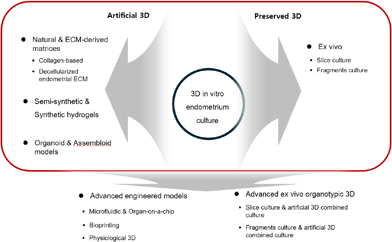
Placental mammalian uterine endometrium, including human endometrium, is a very dynamic reproductive tract and undergoes cyclic proliferation, differentiation, degradation, and sloughing, and regeneration under the coordinated regulation of ovarian sex steroid hormones. These processes are accomplished through continuous remodeling via proliferation, differentiation, cell death, and ECM remodeling. Although the developed 3D culture systems have been used as powerful experimental platforms for studying, there are many limitations in accessing in vivo phenomena. Therefore, this review aims to summarize the major 3D in vitro models developed to recapitulate the human endometrium from the perspectives of ECM- and hydrogel-based models, endometrial organoids and assembloids, engineered or preserved models (Fig. 1). In addition, these were discussed how these models have been applied to the study of hormonal responses and decidualization, implantation, and trophoblast–endometrium interactions, as well as their current limitations and future perspectives for developing physiologically relevant and reproducible 3D endometrial models.
EXTRACELLULAR MATRIX-DERIVED AND HYDROGEL-BASED MODELS
The endometrium is a tissue in which multiple types of endometrial cells interact within the ECM, and therefore, simple 2D cultures have limitations in recapitulating the actual tissue environment (Cook et al., 2017). Hydrogel-based models provide an ECM-like microenvironment and biophysical environment in which cells can grow 3D and interact with the surrounding matrix, making them a useful approach for more physiologically recapitulating the endometrial microenvironment in vitro (Schutte & Taylor, 2012; Cook et al., 2017; Gnecco et al., 2023).
Matrigel is a gelatinous protein mixture, solubilized basement membrane extracts rich in laminin, collagen IV, nidogen/entactin, and heparan sulfate proteoglycans. Its characteristics depend on its components and have been used in both cell culture and in vivo (Passaniti et al., 2022). It can form a hydrogel at physiological temperature and is used for building a 3D microenvironment that supports epithelial survival, polarization, proliferation, and self-organization. Bentin-Ley et al. (1994) successfully cultured the monolayer epithelial cell on Matrigel covered collagen stromal insert. Major advances were made by Turco et al. (2017) and Boretto et al. (2017). Their groups independently established long-term expandable endometrial epithelial organoids (EEOs). Patient-derived organoids have been generated from endometriosis, endometrial hyperplasia, and endometrial cancer (Boretto et al., 2019). Nevertheless, conventional Matrigel-based endometrial organoids have limitations in physiological studies. Matrigel does not accurately reproduce the heterogeneous ECM of the endometrial stroma (Gnecco et al., 2023). Such limitations have stimulated the development of more physiologically defined artificial 3D endometrial models (Table 1).
| Variables | Model | Matrix/Scaffold | Cell components | Main functional application |
|---|---|---|---|---|
| Bentin-Ley et al. (1994) | Collagen-based | Collagen matrix+Matrigel basement-membrane material | Primary endometrial epithelial+stromal cells | Endometrial architecture/epithelial differentiation |
| Abbas et al. (2020) | Collagen-based | Type I collagen porous scaffold | Primary stromal cells+organoid-derived epithelial cells | Hormone response/tissue architecture |
| Jamaluddin et al. (2022) | dECM | Human- and bovine-endometrium-derived dECM hydrogels | Human/mouse endometrial organoids | Organoid culture/disease modeling |
| Gnecco et al. (2023) | Synthetic ECM | Fully synthetic PEG-based hydrogel | Primary epithelial organoids+stromal cells | Hormonal cycling/epithelial–stromal crosstalk |
| Rawlings et al. (2021) | Assembloid | Collagen I hydrogel | Gland-like epithelial organoids+primary stromal cells+human blastocysts | Decidualization/embryo implantation |
| Ahn et al. (2021) | Microfluidic | Multichannel microfluidic 3D platform | Epithelial cell line (Ishikawa)+CRL-4003 stromal fibroblasts+HUVECs | Hormonal response/vasculo-angiogenesis/drug testing |
| Catane et al. (2025) | 3D bioprinting | Alginate-based bioink | Epithelial cell line (RL95-2)+Stromal cell line (THESC), JAR spheroids | Hormonal response/implantation modeling |
3D, three-dimensional; dECM, endometrial extracellular matrix; THESC, telomerase-immortalized human endometrial stromal cell.
Collagen is the most abundant protein family and a major structural component of the ECM, playing important roles not only in providing mechanical support to tissues but also in regulating cell adhesion, migration, and various cellular functions (Gelse et al., 2003; Frantz et al., 2010). In early 3D endometrial models, attempts were made to reconstruct the stromal compartment of the endometrium using collagen, one of the major components of the native ECM. Bentin-Ley et al. (1994) cultured endometrial stromal cells (EnSCs) within a collagen matrix and seeded epithelial cells on top to reconstruct stromal–epithelial architecture, demonstrating that the epithelial cells exhibited characteristics such as polarized columnar morphology, microvilli, and cilia.
Collagen-based models subsequently progressed from simple hydrogel embedding toward more functional tissue-engineering platforms. Schutte & Taylor (2012) encapsulated telomerase-immortalized human endometrial stromal cells (THESCs) within a type I collagen hydrogel and analyzed secretory differentiation, decidualization, and menstrual-like remodeling following hormone withdrawal. Furthermore, Abbas et al. (2020) cultured stromal cells within a porous scaffold fabricated from type I collagen and seeded endometrial organoid fragments onto the surface, thereby simultaneously establishing a luminal-like epithelial layer and an underlying stromal compartment. In this model, both epithelial polarization and the hormone responsiveness of epithelial and stromal cells were maintained, demonstrating that collagen-based matrices can serve not only as structural support but also as scaffolds for recapitulating multicellular endometrial architecture and function.
Collagen-based matrices are biocompatible and can provide a 3D culture environment; however, they have limitations in fully recapitulating the complex biochemical composition of the native endometrial ECM. To overcome these limitations, dECM has been developed to preserve the diverse ECM components and tissue-specific biochemical cues of native endometrial tissue (López-Martínez et al., 2021).
Francés-Herrero et al. (2021) applied endometrial ECM (EndoECM) obtained by decellularizing porcine endometrium to the culture of human endometrial organoids and demonstrated that endometrium-derived ECM could support the growth of endometrial cells. Subsequently, Jamaluddin et al. (2022) generated endometrium-derived ECM hydrogels by decellularizing human and bovine endometrial tissues and applied them to endometrial organoid culture. These hydrogels supported culture at a level comparable to Matrigel and were reported to have the potential to reflect human endometrial biology and pathologies. More recently, hybrid hydrogels combining EndoECM with synthetic components have been developed to improve mechanical properties while maintaining the tissue-specific biochemical cues provided by EndoECM. This approach supports the 3D growth and differentiation of organoids, suggesting the potential to more physiologically recapitulate the native endometrial microenvironment (Gómez-Álvarez et al., 2024).
Natural and ECM-derived hydrogels have the advantage of providing tissue-specific biochemical cues (López-Martínez et al., 2021), however, their biochemical composition and physical properties are intrinsically linked to the source tissue and matrix self-assembly, limiting the independent control of these properties (López-Martínez et al., 2021; Gómez-Álvarez et al., 2024). To overcome these limitations, semi-synthetic hydrogels, which allow the modulation of material properties while maintaining biological activity, and fully synthetic hydrogels, which enable more precise design of their components and mechanical properties, have been applied to 3D endometrial culture.
A representative example of a semi-synthetic hydrogel is gelatin methacryloyl (GelMA), a photo-crosslinkable biopolymer generated by introducing methacryloyl groups into gelatin. Salisbury et al. (2024) cultured primary human EnSC and epithelial gland organoids in GelMA and reported that modulation of the mechanical and biochemical properties of the GelMA hydrogel enabled the growth and maintenance of epithelial gland organoids. Stromal cells encapsulated within GelMA also exhibited responses to hormonal treatment following the induction of decidualization (Salisbury et al., 2024). Zambuto et al. (2024) co-cultured human endometrial microvascular endothelial cells (HEMECs) and stromal cells within GelMA hydrogels to establish a 3D microvascular network and examined changes in vascular network morphology and the secretome according to decidualization status.
Fully synthetic hydrogels have the advantage of providing a chemically defined and tunable microenvironment for 3D culture by enabling the independent and precise control of matrix stiffness, degradability, and cell-adhesion cues (Hernandez-Gordillo et al., 2020). Cook et al. (2017) established a co-culture model system using a PEG-based synthetic modular ECM, in which EnSC encapsulated within the hydrogel and epithelial cells were seeded on top. The matrix was designed to support cell attachment through integrin-binding ligands while also incorporating ECM-binding ligands, thereby enabling the local accumulation of basement membrane components derived from epithelial cells and fibrillar collagen produced by stromal cells, as well as matrix remodeling, and demonstrating a novel application of an ECM-mimicking synthetic hydrogel. Subsequently, Gnecco et al. (2023) expanded this synthetic ECM approach by developing a PEG-based hydrogel containing the collagen-derived adhesion peptide GFOGER, the fibronectin-derived peptide PHSRN-K-RGD, and MMP-cleavable crosslinkers, allowing properties such as stiffness and ligand composition to be controlled. This matrix supported the long-term 3D culture (>15 days) of primary EEOs and stromal cells, and the epithelial and stromal compartments exhibited morphological and molecular responses under different hormonal conditions, demonstrating its potential as a controlled 3D platform for studying cell–cell crosstalk.
ENDOMETRIAL ORGANOIDS AND ASSEMBLOID MODELS
Conventional 2D culture systems have limitations in maintaining primary endometrial epithelial cells over the long term while preserving their tissue-specific morphology and function. To overcome these limitations, organoid culture systems based on the self-organization of primary endometrial epithelium were developed. In 2017, Turco et al. and Boretto et al. independently established long-term expandable EEOs from human endometrial tissue and demonstrated that the generated organoids maintained phenotypes similar to native endometrial glands and responded to reproductive hormones, allowing cycle-associated changes to be recapitulated in organoid culture (Boretto et al., 2017; Turco et al., 2017).
However, these early endometrial organoids primarily recapitulated the glandular epithelial compartment and therefore had limitations in fully reflecting interactions among the different cell types that are important in the native endometrium (Boretto et al., 2017). To overcome this limitation, endometrial assembloids were developed by integrating epithelial organoids with other cell types that constitute the endometrium within a single 3D system. Rawlings et al. (2021) established an assembloid by combining gland-like organoids with primary EnSCs and generated an assembloid model resembling the midluteal endometrium following hormonal induction of decidualization. In addition, co-culture with human blastocysts suggested the potential of this model for studying implantation-associated endometrial responses. Subsequently, Tian et al. (2023) introduced an air–liquid interface culture system to overcome the limitation that conventional organoids and assembloids do not sufficiently recapitulate the luminal epithelium of the native endometrium. Using this approach, they established a more structurally organized human endometrial assembloid containing both luminal epithelium-like and glandular epithelium-like structures (Tian et al., 2023).
ADVANCED ENGINEERED MODELS
To precisely control continuous fluid flow and shear stress, molecular transport between cellular compartments, and the hormonal microenvironment over time, endometrium-on-a-chip models based on microfluidic technology have been developed. Gnecco et al. (2017) compartmentalized and co-cultured human endometrial stromal fibroblasts and endothelial cells in a dual-chamber microfluidic device separated by a porous membrane and applied temporal hormonal stimulation to mimic a 28-day menstrual cycle. In this model, stromal decidualization was maintained, and microfluidic flow-induced shear stress affected endothelial cell cytoskeletal organization and tight-junction formation, demonstrating that a dynamic biophysical environment could be introduced into an endometrial model. Subsequently, Ahn et al. (2021) established a 3D microengineered vascularized endometrium-on-a-chip integrating epithelial, stromal, and vascular compartments. This model spatially organized endometrial epithelial cells, stromal fibroblasts, and endothelial cells to mimic the multilayer architecture of the native endometrium and recapitulated endometrial vasculo-angiogenesis and proliferative- and secretory-phase-associated responses following sex steroid hormone stimulation.
More recently, organ-on-a-chip technology has been extended toward patient-specific models. Lee et al. (2025) developed a vascularized endometrium-on-a-chip integrating patient-derived EEOs with stromal and endothelial cells and demonstrated its potential as a translational platform for evaluating individual endometrial microenvironments and receptivity.
Whereas microfluidic systems have strengths in recreating dynamic biochemical and biophysical environments, 3D bioprinting provides an approach for more precisely recapitulating the structural complexity of tissues by arranging multiple cell types and biomaterials in predefined spatial organizations. Early studies on endometrial bioprinting primarily focused on the development of tissue-engineered constructs for the regeneration of damaged endometrium (Nie et al., 2023).
More recently, Catane et al. (2025) established a hormone-responsive human endometrial bilayer model composed of epithelial and stromal compartments using 3D bioprinting. This model was designed to simultaneously recapitulate the spatial organization of the endometrium and hormone-regulated cellular responses, demonstrating that 3D bioprinting can be utilized not only in regenerative medicine but also as an advanced in vitro endometrial platform for studying embryo implantation.
FUNCTIONAL APPLICATIONS OF THREE-DIMENSIONAL ENDOMETRIAL MODELS
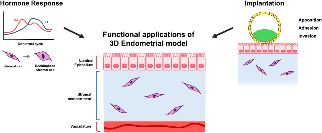
During the menstrual cycle, the endometrium undergoes repeated epithelial proliferation and differentiation, stromal decidualization, and tissue breakdown and regeneration in response to changes in estrogen (E) and progesterone. Therefore, hormone responsiveness in 3D endometrial models is an important functional criterion for evaluating how well these models recapitulate cellular differentiation, multicellular signaling, and cyclic tissue responses observed in the native endometrium, beyond simply expressing hormone receptors.
Artificial 3D endometrial models have been utilized to investigate the mechanisms of hormone-dependent epithelial differentiation. Haider et al. (2019) used human EEOs to analyze how E regulates epithelial cell fate. E stimulation increased the expression of progesterone receptors (PRs) and induced the expression of genes associated with ciliary formation, while also increasing the formation of multiciliated epithelial cells within the organoids. In addition, they demonstrated that the interaction between E and NOTCH signaling regulates endometrial epithelial ciliogenesis, suggesting that endometrial organoids can serve as a functional platform for studying hormone-dependent epithelial differentiation.
Approaches have also been developed to mimic hormonal stimulation more closely resembling the actual menstrual cycle. Olalekan et al. (2017) recellularized decellularized human endometrial scaffolds with primary endometrial cells and subsequently applied a hormone treatment mimicking a 28-day menstrual cycle. The recellularized endometrial tissue-maintained estrogen receptors (ERs) and PRs and secreted prolactin and IGFBP-1 following decidualization stimulation, demonstrating that decidualization responses could be reproduced even under a long-term cyclic hormonal environment. More recently, artificial 3D models have been used to understand decidualization not as an independent response of stromal cells, but as a consequence of epithelial–stromal signaling. Fitzgerald et al. (2023) demonstrated that the basolateral secretome of human EEOs changed in response to steroid hormone and PGE2 stimulation. These findings suggest that endometrial glands act as hormone-responsive secretory compartments that regulate stromal decidualization through paracrine signaling and that organoid models can be used to functionally investigate epithelial–stromal crosstalk. Furthermore, models have recently been developed to reproduce the overall cyclic endometrial response, including not only hormone-dependent differentiation but also menstruation and regeneration. Nikolakopoulou et al. (2026) established an in vitro menstrual cycle by applying sequential E and progesterone stimulation, hormone withdrawal, and tissue disruption to human endometrial organoids. This model recapitulated menstruation-associated epithelial breakdown and regeneration-associated cellular states following secretory differentiation and, in particular, identified a luminal epithelial population and a WNT7A-dependent regenerative response during menstruation and early regeneration. Collectively, these studies demonstrate that organoid endometrial models are evolving beyond simple assessments of hormonal responsiveness to platforms for investigating the mechanisms underlying dynamic cellular differentiation, decidualization, and regeneration throughout the menstrual cycle.
Human embryo implantation is a spatially coordinated process in which the blastocyst apposes and adheres to the receptive endometrial epithelium, followed by trophoblast invasion of the stroma and interaction with the maternal vasculature. Therefore, simple 2D co-culture systems have limitations in fully recapitulating the epithelial barrier, stromal remodeling, and directional trophoblast invasion that occur during implantation. Recent artificial 3D endometrial models have enabled maternal–fetal interactions to be studied in a more physiological environment by directly combining endometrial tissue with trophoblast cells, blastoids, or human embryos (Fig. 2).
Endometrial assembloids have been used to investigate how the functional state of the maternal endometrium influences embryo behavior during implantation. Rawlings et al. (2021) co-cultured decidualized endometrial assembloids with human blastocysts and demonstrated that the presence of senescent decidual stromal cells contributes to the dynamic remodeling of the implantation environment. In assembloids containing senescent decidual cells, migratory decidual cells interacted with expanding blastocysts. In contrast, pharmacological suppression of the senescent decidual cell population accelerated decidualization and generated a robust, static decidual matrix in which non-expanding embryos became entrapped. These findings indicate that implantation is regulated not only by embryo behavior but also by the dynamic remodeling state of the maternal endometrium.
Assembloid engineering technologies have also evolved into models that enable direct observation of trophoblast invasion and maternal vascular interactions. Park et al. (2022) established an implantation-on-a-chip containing primary human extravillous trophoblasts and microengineered maternal vessels, recapitulating the directional migration of trophoblasts toward maternal vessels and their interactions with endothelial cells. This study demonstrated that a microphysiological system can serve as a platform for spatially resolving trophoblast invasion and vascular remodeling following implantation.
More recently, artificial 3D models have been developed to recapitulate the embryo–endometrium interface itself, beyond individual processes of implantation. Shibata et al. (2024) established an apical-out endometrial organoid containing an exposed apical epithelium, stromal cells, and a self-organized endothelial network and co-cultured it with human embryonic stem cell-derived blastoids. This model sequentially recapitulated the early stages of implantation, including apposition, adhesion, and invasion, and demonstrated the process by which syncytial trophoblasts crossed the endometrial epithelial barrier, invaded the stromal compartment, and interacted with stromal cells. Some of these phenomena were also confirmed in experiments with human blastocysts, suggesting the potential of artificial 3D endometrial models to study direct cellular interactions between the embryo and maternal tissue.
Furthermore, Molè et al. (2026) established a human endometrial model with luminal, glandular, and stromal compartments that recapitulated the implantation and early post-implantation development of human embryos and blastoids. Single-cell transcriptomic analysis identified various ligand–receptor interactions at the embryo–endometrium interface, and disruption of a specific signaling interaction between extravillous trophoblasts and EnSC reduced trophoblast outgrowth. These findings demonstrate that recent 3D implantation models are progressing beyond morphological recapitulation toward platforms that enable functional analysis of the molecular mechanisms underlying maternal–fetal crosstalk.
On the other hand, ex vivo systems are considered temporal systems for studying complex in vivo phenomena by discarding the system factors (Schäfer et al., 2011; Dimova et al., 2025). Recently, long-term cultures of authentic and model embryos have become available, particularly in stem cell-based models (Aghajanova, 2020; Yu et al., 2021; Liu et al., 2023; Oldak et al., 2023; Weatherbee et al., 2023). However, to replicate embryo implantation and related diseases, there is a limitation in the decidual uterus, which has natural complexity. As mentioned, artificial 3D systems aim to mimic the physiological environment of in vivo organs. Based on that point, ex vivo use of tissue slices or fragments retains the original complexity. It means that the ex vivo system is useful for implantation studies with the extended artificial 3D system. Recently, Hiraoka and Ikawa’s group suggested the usefulness of ex vivo culture of endometrial fragments for studying embryo implantation (Hiraoka et al., 2025). It means that innovations in culture systems enhance the usefulness of artificial 3D and ex vivo native-preserved 3D systems.
CURRENT LIMITATIONS AND FUTURE PERSPECTIVES
Current artificial 3D endometrial models are progressively recapitulating the structure and function of the native endometrium through diverse approaches, including hydrogels, organoids, assembloids, and organ-on-a-chip systems. However, each model has strengths in specific aspects, such as tissue-specific ECM, epithelial self-organization, multicellular interactions, or dynamic microenvironments, and limitations remain in simultaneously recapitulating the complex cellular composition and spatial organization of the native endometrium within a single system (Agustina-Hernández et al., 2024). In addition, standardization of culture conditions and physiological consistency remain challenges in 3D models, while microfluidic and organ-on-a-chip systems have limitations in terms of technical complexity and accessibility (Agustina-Hernández et al., 2024).
Therefore, future development of artificial 3D endometrial models should consider not only increasing the number of cell types included and the structural complexity but also reproducing the fundamental architecture of native tissue in a consistent, experimentally accessible manner. The human endometrium is spatially organized into the luminal epithelium, glandular epithelium, and underlying stromal compartment, and the absence of an intact luminal epithelium in conventional endometrial organoids and assembloids has been identified as an important limitation in recapitulating endometrial receptivity (Tian et al., 2023). In particular, during implantation, the trophectoderm attaches to the receptive luminal epithelium and subsequently penetrates the epithelial barrier to invade the underlying endometrial tissue (Ruane et al., 2022). Therefore, a stratified tissue model based on a well-defined foundational architecture with an accessible epithelial surface and underlying 3D stroma could serve as a fundamental structural platform for studying sequential trophoblast–endometrium interactions. Ultimately, the development of artificial 3D endometrial models that progressively integrate the biochemical, cellular, spatial, and temporal characteristics of the native endometrium while maintaining reproducibility and accessibility and achieving standardization across models will be an important future direction for studying human endometrial physiology and implantation (Agustina-Hernández et al., 2024). An ex vivo system is also suggested as an excellent model through the improvement of methods. As mentioned, the ex vivo system already has the native preserved 3D structure and function. So, it is very useful when it is improved logically. The combination of artificial 3D culture strategies with ex vivo organotypic 3D systems, or vice versa, can be helpful for studying implantation and physiological abnormalities.
CONCLUSION
The human endometrium has diverse cell populations and the ECM, which are spatially organized and undergo cyclic changes by the menstrual cycle. To recapitulate this complexity in vitro, artificial 3D endometrial models have evolved from early collagen-based matrices and hydrogel systems to tissue-specific dECM, semi-synthetic hydrogels, endometrial organoids and assembloids, and engineered platforms such as microfluidic organ-on-a-chip systems and 3D bioprinting. In addition, ex vivo organotypic native-preserved 3D systems of the endometrium are improved through the systematic development of 3D culture models. Although current artificial 3D models effectively recapitulate specific structural or functional characteristics of the native endometrium, limitations remain in their ability to simultaneously reproduce the biochemical environment, cellular composition, spatial architecture, and temporal hormonal dynamics within a single system (Kleinová et al., 2024). A modular approach, in which the fundamental architecture of native tissue is first reproduced in a stable and well-defined manner and biological components and dynamic cues may provide a practical strategy for future model development. In particular, selectively integrating vascular, immune, and trophoblast components into a reproducible tissue architecture that includes an accessible luminal epithelial surface and an underlying stromal compartment may strike a balance between physiological relevance and experimental controllability. Enhancing ex vivo methods with artificial 3D culture also has synergistic effects on the artificial 3D system (Hiraoka et al., 2025). Ultimately, the development of 3D models that integrate the biochemical, cellular, spatial, and temporal characteristics of the native endometrium while maintaining high reproducibility and accessibility and establishing standardized culture conditions will provide an important foundation for understanding the mechanisms of human endometrial physiology and implantation and for studying reproductive disorders.
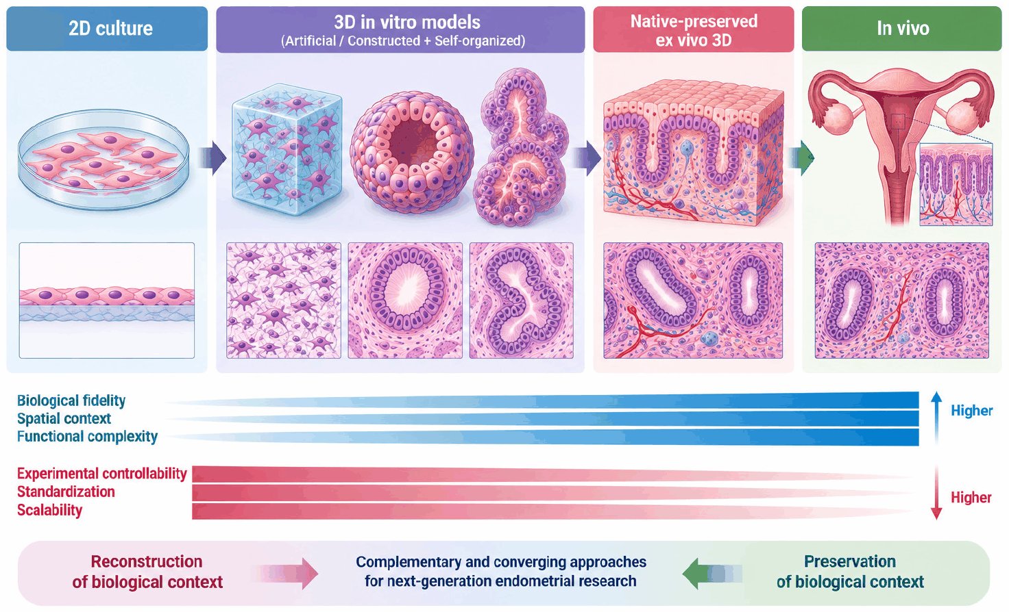
The next generation of 3D culture calls for a conceptual shift from viewing 3D merely as a geometric property toward understanding as a continuum of biological context, spanning 2D culture, artificial/self-organoid 3D, native-preserved ex vivo 3D and in vivo. Along this continuum, engineered models progressively reconstruct selected features of the tissue microenvironment, whereas ex vivo organotypic native preserved 3D models preserve pre-existing native architecture, ECM, cellular heterogeneity, and spatial interactions. Rather than representing a simple hierarchy of model superiority, these systems offer different balances between experimental controllability and biological fidelity (Fig. 3). Future experimental biology may therefore advance through the convergence of engineering-driven reconstruction and native-context preservation, ultimately narrowing the conceptual boundaries between the models.