Development & Reproduction
The Korean Society of Developmental Biology
ARTICLE

Early Gonadal Differentiation of the Protogynous Red Spotted Grouper, Epinephelus akaara

Hyun Kyu Kim1, Jung-Hyun Kim1, Woo Sik Kim1, Hea Ja Baek2, Joon Yeong Kwon†,1
1Dept. of Aquatic Life Medical Science, Sunmoon University, Asan 336-708, Korea
2Dept. of Marine Biology, Pukyong National University, Busan 48513, Korea
Corresponding Author : Joon Yeong Kwon, Dept. of Aquatic Life Medical Science, Sunmoon University, Asan 336-708, Korea. Tel. : +82-41-530-2284, jykwon@sunmoon.ac.kr

Copyright © 2015 © Korean Society of Developmental Biology. All Rights Reserved. This is an Open-Access article distributed under the terms of the Creative Commons Attribution Non-Commercial License (http://creativecommons.org/licenses/by-nc/3.0/) which permits unrestricted non-commercial use, distribution, and reproduction in any medium, provided the original work is properly cited.

Received: Oct 10, 2015 ; Revised: Nov 2, 2015 ; Accepted: Nov 17, 2015

ABSTRACT

Red spotted grouper, Epinephelus akaara is a popular aquaculture species in many Asian countries. This species is a protogynous hermaphrodite that first differentiates into female and changes to male later. Due to this reproductive characteristic, stable supply of male and female gametes is a key to the success of seed production in this species. Thus, understanding early gonadal differentiation is required to develop effective sex control techniques. Red spotted grouper were reared in indoor tanks and sampled every 5 days from 40 days post-hatch (DPH) to 130 DPH. Changes of gonadal tissues were examined and analyzed by means of histology. A pair of gonadal primordium has already existed underneath the kidney in the posterior part of the body cavity at 38 DPH when this study began. Gonadal primordia of 38, 40 DPH consisted of germ cells surrounded by a few somatic cells. The blood vessel was observed in the gonadal primordium at 45 DPH. The number of somatic cells and size of gonadal primordium increased age-dependently up to 60 DPH. Formation of ovarian cavity was obvious by two protuberant aggregations of somatic cells at 65 DPH. Completed ovarian cavity and oogonia were first observed in the gonad of one fish sample at 105 DPH. Based on these histological observations, it can be suggested that induction of primary male differentiation could be more successfully applied at around 60 DPH in this species.


INTRODUCTION

Red spotted grouper, E. akaara is one of the most popular and important grouper species for aquaculture in south-east and east Asian countries including Japan, Taiwan, Hong Kong and Indonesia thanks to its fast growth and high market value. Despite the significant economic value, large scale seed production techniques for this species have not been completely developed to date. One of the major difficulties in seed production is associated with the reproductive characteristics of grouper species.

Groupers that belong to the genus of Epinephelus are known to be protogynous hermaphrodites that first differentiates into female and changes to male later (Tan & Tan, 1974; Brusle- Sicard et al., 1992; Shapiro et al., 1993; Bhandari et al., 2003; Alam & Nakamura, 2007; Murata et al., 2009; Liu & Sadovy, 2009; Sao et al., 2012). Natural sex change of E. akaara from female to male occurs 5 to 6 years old (Li et al., 2006; Li et al., 2007). Brood stocks of E. akaara, however, often show male preponderance within a few years under artificial conditions, suggesting bi-directional sex change (Tanaka et al., 1990; Okumura, 2010). These are indicative of poor understanding on the process of sex differentiation and sex reversal in this species.

In many fish, hormonal treatment can induce functional sex change from female to male as well as produce allmale or all-female population of young fish (Yamazaki, 1983; Hunter & Donaldson, 1983). Red spotted grouper is one of those species that requires the help of hormonal treatment to achieve stable supply of male and female gametes for successful seed production. Tanaka et al. (1990) have found male fish in 1 year old group of this species, implying the possibility of primary male differentiation. However, hormonal induction of primary male differentiation in this species has not been satisfactory so far (Lee et al., 2014). Understanding on the process of early gonadal differentiation would improve sex reversal technique for this species. Therefore, we investigated early development of gonad for more than 3 months from hatching based on histological changes, and analyzed the relationship between fish size and gonadal differentiation in red spotted grouper, E. akaara.

MATERIAL & METHODS

Red spotted grouper, E. akaara were collected twice from Cheongsol aquaculture farm in Muan-gun, Jeonnam, Korea, and transported to a laboratory in Sunmoon University (38 and 40 DPH, day post hatching). Fish were reared at 27 ± 1°C and 31 ± 1‰ in indoor tanks. Photoperiod was maintained at 14 hours light : 10 hours dark (14L:10D). Fish were fed a commercial diet (40 to 80 DPH: Ottohime, Japan; 80 to 130 DPH: Myungsun, Korea) ad libitum twice a day. Sampling was carried out 5 days between 40 to 105 DPH. Additionally, fish at 38, 120, 130 DPH were sampled further.

These fish were anesthetized by 50 ppm benzocaine (Sigma) and measured for body weight (g) and total length (mm). Whole body samples were first fixed in 10% formalin for about 12 h. After elimination of head and tail from the fixed fish, the remaining tissues were further fixed in 10% formalin again for about 12 h. The fixed tissues were then dehydrated with ascending alcoholic series, cleared in xylene, and embedded in paraffin wax. Tissues were cut to a thickness of 5~8 μm, stained with haematoxylin & eosin and observed under light microscope (DM500, Leica, Germany).

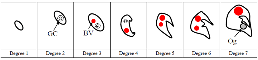
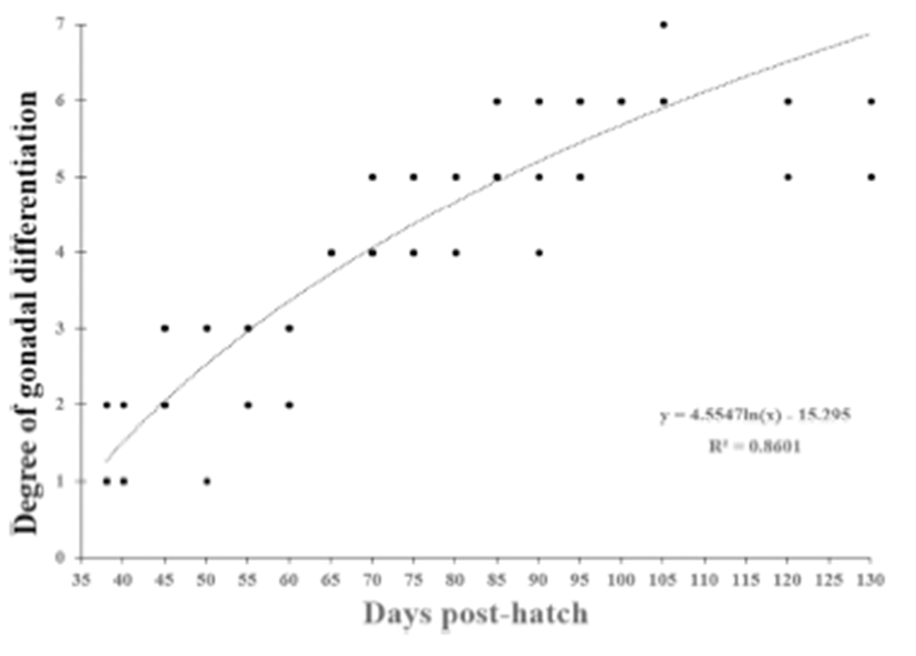
Gonadal differentiation was categorized as undifferentiated gonad phase, differentiated gonad phase and ovarian phase on the basis of two protuberant aggregations of somatic cells in gonadal primordium (onset of ovarian cavity formation) and appearance of ovarian cavity and oogonia (onset of ovarian phase). To analyze correlation between gonadal differentiation degree and fish size, gonadal differentiation degree was further divided into seven steps and granted score one to seven then we carried out linear regression analysis.

RESULTS

1. Fish growth during the early gonadal differentiation

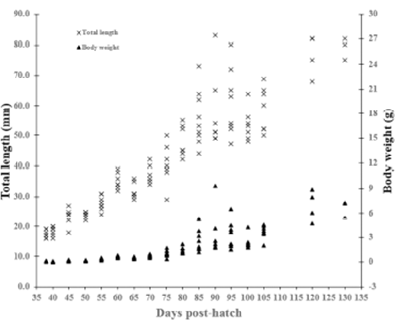
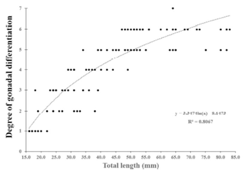
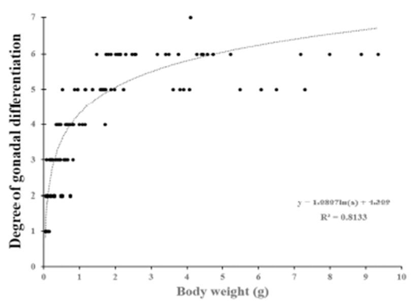
In undifferentiated gonadal phase of red spotted grouper, E. akaara (from 38 to 60 DPH), total length and body weight ranged from 17.4 to 35.1 mm and 0.1 to 0.6 g, respectively. In initially differentiated gonad (65 DPH), total length and body weight were 31.3 ± 0.78 mm and 0.5 ± 0.04 g, respectively. In differentiated gonadal phase (65 to 130 DPH), total length and body weight ranged from 31.3 to 79.0 mm and 0.5 to 6.9 g, respectively (Fig. 1).

dr-19-4-209-g001
Fig. 1. Total length (mm) or body weight (g) of red spotted grouper E. akaara.
Download Original Figure
2. Histology of early gonadal differentiation

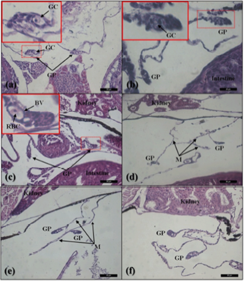
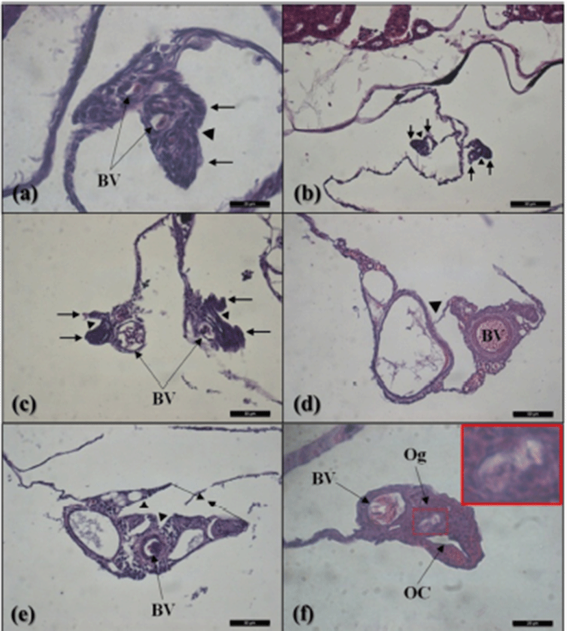
The gonadal primordium of red spotted grouper, E. akaara was observed at 38 DPH (Fig. 2). A pair of gonadal primordium was located in the middle part of mesentery, immediately below the kidney in the body cavity. Gonadal primordium of 38, 40 DPH consisted of germ cells surrounded by a few somatic cells. The blood vessel was observed in the gonadal primordium at 45 DPH. The number of somatic cells and size of gonadal primordium increased age-dependently up to 60 DPH. In the gonadal primordium of all fish at 65 DPH, formation of ovarian cavity was obvious by two protuberant aggregations of somatic cells (Fig. 3). In most of the samples up to 130 DPH, the two elongating walls of somatic cells developed further but not connected to each other to complete the ovarian cavity. However, completed ovarian cavity and oogonia were first observed in the gonad of one fish sample at 105 DPH.

dr-19-4-209-g002
Fig. 2. Undifferentiated gonad in red spotted grouper at 38 to 60 DPH. (a, b) Gonadal primordium of 38, 40 DPH consisted of germ cells surrounded by a few somatic cells. Scale bar (a) = 50 μm, Scale bar (b) = 20 μm; (c) Blood vessels with red blood cells were first appeared at 45 DPH within the gonadal primordium. Scale bar = 50 μm; (d, e, f) The number of somatic cells and size of gonadal primordium at 50, 55 and 60 DPH increased age-dependently. Scale bar (d, e, f) = 50 μm. GC: germ cell; GP: gonadal primordium; BV: blood vessel; RBC: red blood cell; M: mesentery.
Download Original Figure
dr-19-4-209-g003
Fig. 3. Formation process of ovarian cavity in red spotted grouper at 65 to 105 DPH. (a) Gonadal primordium at 65 DPH showed morphological characteristic of the onset of ovarian cavity formation by two protuberant aggregations of somatic cells in gonadal primordium. Scale bar = 20 μm; (b) Two aggregations of somatic cells further developed in gonadal primordium at 70 DPH. Scale bar = 50 μm; (c, d, e) Gonadal primordium at 80, 90, 100 DPH during ovarian cavity formation. Scale bar (c, d, e) = 50 μm; (f) Formation of completed ovarian cavity at 105 DPH. Scale bar = 20 μm; BV: blood vessel; Og: oogonia; OC: ovarian cavity; ▲: Formation of ovarian cavity; Black head arrow: aggregations of somatic cells.
Download Original Figure
3. Correlation between fish size and the degree of gonadal differentiation

We divided the degree of gonadal differentiation into seven (Fig. 4). The correlation of gonadal differentiation degree with DPH was positive and statistically significant (R2 = 0.8607, P < 0.05) (Fig. 5). However, correlation between fish size (total length and body weight) and the degree of gonadal differentiation were not statistically associated (P > 0.05) (Fig. 6 and 7).

dr-19-4-209-g004
Fig. 4. Degrees of gonadal differentiation from gonadal primordium into ovarian phase in red spotted grouper E. akaara. Degree 1: gonadal primordium without any prominent germ cells (GC); Degree 2: appearance of GC surrounded by a few somatic cells; Degree 3: Blood vessels with red blood cells are first appeared; Degree 4: the onset of ovarian cavity formation by two protuberant aggregations of somatic cells in gonadal primordium; Degree 5: the number of somatic cells and size get increased; Degree 6: the ends of two aggregations of somatic cells further developed and get closed; Degree 7: completion of ovarian cavity formation and first appearance of oogonium (Og).
Download Original Figure
dr-19-4-209-g005
Fig. 5. Relationship between degrees of gonadal differrentiation and days post-hatch (DPH) in red spotted grouper E. akaara.
Download Original Figure
dr-19-4-209-g006
Fig. 6. Relationship between degrees of gonadal differrentiation and total length (mm) in red spotted grouper E. akaara.
Download Original Figure
dr-19-4-209-g007
Fig. 7. Relationship between degrees of gonadal differrentiation and body weight (g) in red spotted grouper E. akaara.
Download Original Figure

DISCUSSION

1. First appearance of gonadal primordium and germ cell

A pair of gonadal primordium has already existed underneath the kidney in the posterior part of the body cavity at 38 DPH when this study began. In other grouper species, appearance of gonadal primordium was first observed at 20 DPH in longtooth grouper, E. bruneus; at 18 DPH in malabar grouper, E. malabaricus; at 3 weeks after hatching (WAH) in orange spotted grouper, E. coioides (Murata et al., 2009; Liu & Sadovy, 2009; Sao et al., 2012). This result indicates that the first appearance of gonadal primordium in red spotted grouper could be much earlier than 38 DPH.

Germ cells as well as gonadal primordium were found at 38 DPH in this species. The timing of germ cell appearance in grouper depends on fish species (Murata et al., 2009; Liu & Sadovy, 2009; Sao et al., 2012). The appearance of germ cell was found at 40 to 50 DPH in longtooth grouper, E. bruneus; at 39 DPH in malabar grouper, E. malabaricus; at 7 WAH in orange spotted grouper, E. coioides. Appearance of germ cell in red spotted grouper seems to be earlier than other grouper species mentioned here.

2. Formation of ovarian cavity

Morphological characteristic of ovarian cavity formation was first observed in gonadal primordium of all fish at 65 DPH in this species. This morphological characteristic is similar to what occurs in the onset of ovarian cavity formation in other teleost fish such as Cottus bairdii, Oreochromis aureus, O. mossambicus, Carassius auratus, O. niloticus (Hann, 1927; Eckstein & Spira, 1965; Nakamura, 1978; Nakamura & Nagahama, 1985). The morphological characteristic was also very similar to other grouper species such as malabar grouper, E. malabaricus; longtooth grouper, E. bruneus; orange spotted grouper, E. coioides (Murata et al., 2009; Liu & Sadovy, 2009; Sao et al., 2012).

In the present study, completed ovarian cavity and oogonia were first observed in the gonad of one fish sample at 105 DPH. Nakamura et al. (1998) reported that physiological sex determination in germ cells occurs in synchrony with gonadal sex differentiation. Appearance of ovarian cavity and oogonia means completion of ovarian sex differentiation. However, ovarian cavity was not completed in many fish until 130 DPH. In other grouper, the ovarian cavity formation was reported to be completed at 110 to 130 DPH in longtooth grouper, E. bruneus and at 144 DPH in malabar grouper, E. malabaricus (Murata et al., 2009; Sao et al., 2012). Liu & Sadovy, (2009) have also shown that the completed ovarian cavity was first observed in orange spotted grouper, E. coioides and humpback grouper, Cromoleptes altivelis at 16 to 30 WAH and at 22 WAH respectively.

3. Correlation between fish size and the degree of gonadal differentiation

The correlation of gonadal differentiation degree with DPH was statistically significant while correlations of gonadal differentiation degree with fish size (total length and body weight) were not. It could be expected that the bigger fish may have more differentiated gonad than the smaller fish at the same age. However, analysis from the present study suggests that the progress of gonadal differentiation depends on the age of fish (DPH) rather than the size of fish in this species at least until the completion of ovarian cavity.

4. Protogynous hermaphroditism

The gonads of all fish examined in this studied differentiated into ovarian structure. This confirms red spotted grouper is protogynous species, supporting previous studies (Sadovy de Mitcheson & Liu, 2008). Serranidae hermaphrodites are classified as Serranus type, Rypticus - Anthias type and Epinephelus type depending on gonadal development and sex change type (Smith, 1965). Sexual patterns of red spotted grouper, E. akaara belong to Epinephelus genus which is protogynous hermaphrodites with bidirectional sex change (Sadovy de Mitcheson & Liu, 2008). Protogynous hermaphroditism is divided into monandry and diandry (Reinboth, 1967). However, protogynous hermaphroditism in red spotted grouper is yet to be further clarified.

5. Suggestion for artificial sex change

In fish, the induction of artificial sex change is most effective during gonadal sex differentiation (Yamamoto, 1969; Nakamura & Takahashi, 1973; Nakamura et al., 1998). Lee et al. (2014) have shown that induction of primary male differentiation can be possible by a treatment with 17α-methyltestosterone in red spotted grouper at 70 DPH. In the present study, undifferentiated gonadal phase was up to 60 DPH, and ovarian differentiation began between at 60 to 65 DPH. Based on this result, it can be suggested that induction of primary male differentiation could be more successfully applied at around 60 DPH in this species.

ACKNOWLEDGEMENT

This research was supported by Golden Seed Project, Ministry of Agriculture, Food and Rural Affairs (MAFRA), Ministry of Oceans and Fisheries (MOF), Rural Development Administration (RDA) and Korea Forest Service (KFS) (213004-04-03-SB620).

REFERENCES

1.

Alam MA, Nakamura M. Efferent duct differrentiation during female-to-male sexchange in honeycomb grouper, Epinephelus merra. J Fish Biol. 2007; 71 p. 1192-1202.

2.

Bhandari RK, Komuro H, Nakamura S, Higa M, Nakamura M. Gonadal restructuring and correlative steroid hormone profiles during natural sex change in protogynous honeycomb grouper (Epinephelus merra). Zool Sci. 2003; 20 p. 1399-1404.

3.

Brusle-Sicard S, Debas L, Fourcault B, Fuchs J. Ultrastructural study of sex inversion in a protogynous hermaphrodite, Epinephelus microdon (Teleostei, Serranidae). Reprod Nutr Dev. 1992; 32 p. 393-406.

4.

Eckstein B, Spira M. Effect of sex hormones on gonadal differentiation in a cichlid, Tilapia aurea. Biol Bull. 1965; 129 p. 482-489.

5.

Hann HW. The history of the germ cells of Cottus bairdie. Girard J Morphol Physiol. 1927; 43 p. 427-498.

6.

Hunter GA, Donaldson EM. In: Hoar WS, Randall DJ, Donaldson EM, editors. Hormonal sex control and its application to fish culture. Fish Physiology. 1983; Academic PressNew York9B p. 223-303.

7.

Lee CH, Hur SW, Na OS, Baek HJ, Noh CH, Han SH, Lee YD. Induction of primary male in juvenile red spotted grouper Epinephelus akaara by immersion of 17α-methyltestosterone. Dev Reprod. 2014; 18 p. 127-131.

8.

Li GL, Liu XC, Lin HR. Effects of aromatizable and nonaromatizable androgens on the sex inversion of redspotted grouper (Epinephelus akaara). Fish Physiol Biochem. 2006; 32 p. 25-33.

9.

Li GL, Liu XC, Lin HR. Seasonal changes of serum sex steroids concentration and aromatase activity of gonad and brain in red spotted grouper (Epinephelus akaara). Ani Reprod Sci. 2007; 99 p. 156-166.

10.

Liu M, Sadovy de Mitcheson Y. In: Liao IC, Leaño EM, editors. Chapter 7: Grouper mariculture in mainland China and Hong Kong. The Aquaculture of Groupers. 2008; Asian Fisheries SocietyManila, PhilippinesWorld Aquaculture SocietyLouisiana, USAThe Fisheries Society of TaiwanKeelungTaiwan and National Taiwan Ocean UniversityKeelung, Taiwan p. 111-142.

11.

Liu M, Sandovy Y. Gonad development during sexual differentiation in hatchery-produced orange-spotted grouper (Epinephelus coioides) and humpback grouper (Cromileptes altivelis) (Pisces: Serrannidae, Epinephelinae). Aquacult. 2009; 287 p. 191-202.

12.

Murata R, Karimata H, Alam MA, Nakamura M. Gonad sex differentiation in the malabar grouper (Epinephelus malabaricus). Aquacult. 2009; 293 p. 286-289.

13.

Nakamura M. Morphological and experimental studies on sex differentiation of the gonad in several teleost fishes. Ph.D. Thesis. Hokkaido University. Hokkaido. Japan. 1978.

14.

Nakamura M, Kobayashi T, Chang XT, Nagahama Y. Gonadal sex differentiation in teleost fish. J Exp Zool. 1998; 281 p. 362-372.

15.

Nakamura M, Nagahama Y. Steroid producing cells during ovarian differentiation of the tilapia Sarotherodon niloticus. Dev Growth Differ. 1985; 27 p. 701-708.

16.

Nakamura M, Takahashi H. Gonadal sex differentiation in tilapia, with special regard to the time of estrogen treatment effective in inducing complete feminization of genetic males. Bull Fac Hokkaido Univ. 1973; 24 p. 1-13.

17.

Okumura S. Evidence of sex reversal towards both directions in reared red spotted grouper Epinephelus akaara. Fish Sci. 2001; 67 p. 535-537.

18.

Reinboth R. Biandric teleost species. Gen Comp Endocrinol. 1967; 9-486.

19.

Sao PN, Hur SW, Lee CH, Lee YD. Gonadal sex differentiation of hatchery-reared longtooth grouper (Epinephelus bruneus). Dev Reprod. 2012; 16 p. 185-193.

20.

Shapiro DY, Sadovy Y, McGehee MA. Periodicity of sex change and reproduction in the red hind, Epinephelus guttatus, a protogynous grouper. Bull Mar Sci. 1993; 53 p. 1151-1162.

21.

Smith LC. The patterns of sexuality and the classifycation of serranid fishes. Amer Mus Nov. 1965; 2007 p. 1-20.

22.

Tan SM, Tan KS. Biology of the tropical grouper, Epinephelus tauvina (Forskal). I: A preliminary study on hermaphroditism in E. tauvina. Singap J Prim Ind. 1974; 2 p. 123-133.

23.

Tanaka H, Hirose K, Nogami K, Hattori K, Ishibashi N. Sexual maturation and sex reversal in red spotted grouper, Epinephelus akaara. Bull Natl Res Inst Aquacult. 1990; 17 p. 1-15.

24.

Yamamoto T. In: Hoar WS, Randall DJ, editors. Sex differentiation. Fish Physiology. 1969; Academic PressNew YorkIII p. 117-175.

25.

Yamazaki F. Sex control and manipulation in fish. Aquacult. 1983; 33 p. 329-354.