Development & Reproduction
Korean Society of Developmental Biology
Research articles

Gonadal Abnormality and Intersexuality of Oplegnathus fasciatus (Teleostei: Oplegnathidae) Collected from the Southern Coast of Korea: A Case Report

Ji Sung Choi1https://orcid.org/0000-0002-8312-795X, So Ryung Shin1https://orcid.org/0000-0002-2888-1378, Hyeon Jin Kim1https://orcid.org/0000-0001-9208-4616, Hyejin Kim2https://orcid.org/0000-0003-3853-0034, Yeonghye Kim3https://orcid.org/0000-0002-5927-0641, Jung Sick Lee1,https://orcid.org/0000-0001-6292-328X
1Dept. of Aqualife Medicine, Chonnam National University, Yeosu 59626, Korea
2Fisheries Seed and Breeding Research Institute, National Institute of Fisheries Science, Haenam 59002, Korea
3Fisheries Resources Management Division, National Institute of Fisheries Science, Busan 46083, Korea
Corresponding author Jung Sick Lee Dept. of Aqualife Medicine, Chonnam National University, Yeosu 59626, Korea Tel: +82-61-659-7172 Fax: +82-61-659-7179 E-mail: ljs@jnu.ac.kr

Copyright © 2021 The Korean Society of Developmental Biology. This is an Open Access article distributed under the terms of the Creative Commons Attribution Non-Commercial License (http://creativecommons.org/licenses/by-nc/4.0/) which permits unrestricted non-commercial use, distribution, and reproduction in any medium, provided the original work is properly cited.

Received: May 06, 2021 ; Revised: Jul 21, 2021 ; Accepted: Aug 26, 2021

Published Online: Sep 30, 2021

Abstract

We report on the gonadal structure abnormality and intersexuality found from a survey regarding the reproductive potential of the rock bream, Oplegnathus fasciatus, along the southern coast of Korea. In total, 448 samples were collected for histological analysis (total length 27.5±6.8 cm; total weight 522.9±385.6 g). The sex ratio (F:M) was 1:0.46 (n=307:141), with 68.5% being females. The frequency of oocyte atresia and ovarian neoplasm in the females was 85.3% and 21.5%, respectively, and the frequency of testicular lobule and cyst deformation and testicular neoplasm in the males was 73.1% and 37.6%, respectively. The scale formation in the ovary and testis was approximately 2.0% for both sexes. The intersexuality was 42.2%, with the intersexuality among females being 49.5%, which was significantly higher than that of males (26.2%). Such findings indicate a negative impact on the reproductive output of the rock bream along the southern coast of Korea; however, a more detailed study is required to ascertain accurate causes.

Keywords: Oplegnathus fasciatus; Gonadal abnormality; Intersexuality

INTRODUCTION

The first phase of the ecological risk assessment procedure due to environmental factors is hazard identification (NRC, 1983). Among the biomarkers used for assessing ecotoxicity, reproductive biomarkers are an integral part of assessing long-term and continuous effects from environmental factors (Huggett et al., 1992; Schmitt et al., 2005). Histological biomarkers, including sex, stages of gonadal development, tumors, intersex, and atresia, are also an essential component for researching the reproductive health of fishes (Blazer, 2002; Kokokiris et al., 2005; Lee et al., 2010; Colin et al., 2016).

The rock bream, Oplegnathus fasciatus, is a teleost that belongs to Perciformes and Oplegnathidae. It is a coastal fish species that inhabits coastal waters of the South China Sea, Japan, and Korea (Kim et al., 2005). In Korea, the rock bream is a very important species for aquaculture and conservation of fishery resources. The rock bream is produced artificially from the hatched larvae to the early stage before the juveniles are grown to approximately 5.0 cm in total length and ready to be released to the wild. Nearly 9 million fishes were released to the wild from 2012 to 2015 (FIRA, 2015). The present study aimed to investigate the gonad structural abnormality and intersexuality of the rock bream, O. fasciatus, inhabiting the southern coast of Korea, measured during a survey of their reproductive potential.

MATERIALS & METHODS

1. Specimens

In total, 448 rock bream were used in the analysis with total length of 27.5±6.8 cm and total weight of 522.9±385.6 g. The samples were collected using stow nets and trawl nets in waters near Sacheon, Mokpo, and Yeosu of Korea (Fig. 1).

2. Histological analysis

Five preparations of 1 cm2 area were analyzed from each individual after categorizing the gonads by fore, middle, and rear parts using the methods described by Lee et al. (2010). Each sample was fixed with 10% neutral buffered formalin and then dehydrated through an ethanol series (70%–100%). The tissues were embedded in paraplast (McCormick Scientific, St. Louis, MO). Embedded tissues were serially sectioned to a thickness of 4–6 µm using a microtome (RM2235, Leica, Germany). Samples were stained with Mayer’s hematoxylin–0.5% eosin (H-E) stain and Masson’s trichrome stain.

3. Statistical analysis

The chi-square test is conducted with SPSS 21.0 software (SPSS, Chicago, IL, USA) to verify the significant difference in sex ratio (p<0.05).

dr-25-3-123-g1
Fig. 1. Sampling area of the rock bream, Oplegnathus fasciatus.
Download Original Figure

RESULTS AND DISCUSSION

1. Sex ratio

The sex ratio of O. fasciatus was 1:0.46, with 307 female specimens and 141 male specimens. Females made up approximately 68.5% of the entire sample, showing a significantly higher composition than that of the males (p<0.05; Table 1).

Table 1. Sex ratio of the rock bream, Oplegnathus fasciatus
Specimen number Sax ratio Chi-square p-value
Total Female Male F:M F/F+M (%)
448 307 141 1:0.46 68.5 61.509 0.000
Download Excel Table
2. Ovarian structure abnormality
1) Oocyte atresia

As oocyte atresia proceeds, the shape of the oocytes becomes irregular. We observed shrinking of the zona radiata and more eosinophilic reactions than those in the normal oocytes (Fig. 2A and B). Moreover, we identified the granulation of chromatin and the collapse of the nuclear membrane. A loss of eosinophilic yolk granules was observed in the cytoplasm. The yolk granules in the cortex and medulla were homogeneous, with the medulla found to be more basophilic in the H-E stain than the cortex part (Fig. 2B and C). Oocyte atresia occurred in 85.3% of samples (Table 2).

Table 2. Abnormal type and frequency of gonadal structure in the rock bream, Oplegnathus fasciatus
Abnormal type Frequency (%)
Ovary Oocyte atresia (Oa) 85.34 (n=262/307)
Ovarian neoplasia (On) 21.50 (n=66/307)
Scale formation in the ovary (So) 1.95 (n=6/307)
Oa * On 19.22 (n=59/307)
Oa * So 0.65 (n=2/307)
On* So 0.33 (n=1/307)
Oa * On * So 0.33 (n=1/307)
Testis Deformation of testicular lobule and cyst (Dt) 73.05 (n=103/141)
Testicular neoplasia (Tn) 37.59 (n=53/141)
Scale formation in the testis (St) 2.13 (n=3/141)
Dt*Tn 26.95 (n=38/141)
Dt * St 1.42 (n=2/141)
Tn * St (n=0/141)
Dt*Tn*St (n=0/141)
Download Excel Table
dr-25-3-123-g2
Fig. 2. Photomicrographs of oocytes in the rock bream, Oplegnathus fasciatus. (A) Normal vitellogenic oocyte. (B) and (C) Oocyte atresia. H-E stain. N, nucleus; No, nucleolus; Oc, oocytes; Yg, yolk granules; Zr, zona radiata; *, atretic oocytes.
Download Original Figure
2) Ovarian neoplasm

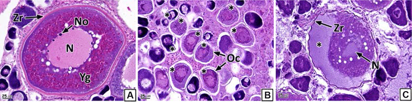
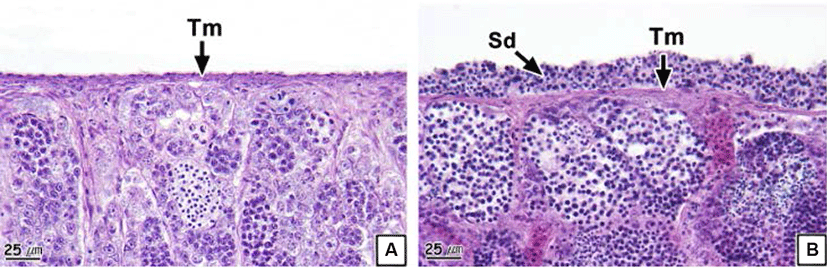
We could not morphologically identify the neoplasm in the form of nodules or tumors from the ovaries. However, histological analysis showed a cluster of multiple oocytes in different stages of development and a newly formed connective tissue layer outside the ovarian membrane (Fig. 3). The newly formed connective tissue layer turned blue in the Masson’s trichrome stain (Fig. 3C). Oocytes and fatty tissues showed eosinophilic stainability and vacuoles in the H-E stain, respectively (Fig. 3B). The prevalence of ovarian neoplasm recorded was 21.5% (Table 2).

dr-25-3-123-g3
Fig. 3. Photomicrographs of ovary in the rock bream, Oplegnathus fasciatus. (A) Normal ovary. (B) and (C) Ovarian neoplasm. (A) and (B) H-E stain, (C) Masson’s trichrome stain. Section showing the formation of numerous oocytes (Oc), connective tissue and fatty tissue (Ft) in the outer ovarian membrane (Om). Ol, ovarian lamella.
Download Original Figure

Oocyte atresia is a normal phenomenon during the degeneration of oocytes after spawning in teleosts (Genten et al., 2009). Oocyte atresia has been reported as an endocrine disorder of female aquatic animals, which is inflicted on oogenesis owing to various environmental factors. The species currently affected include the common carp (Blazer, 2002); sole, Solea solea (Cuevas et al., 2015); brown bullhead, Ameiurus nebulosus; smallmouth bass, Micropterus dolomieu; yellow perch, Perca flavescens (Pinkney et al., 2017) and Mytilus trossulus (Smolarz et al., 2017). The histological properties seen in the rock bream during the present study were similar to those from these previous studies.

Gonadal neoplasm of aquatic animals has been mainly reported in the form of nodules, tumors, or mixed germ cell-stromal neoplasia from crucian carp, Carassius carassius (Fregeneda-Grandes et al., 2010); softshell clam, Mya arenaria (Barber, 1996) and mussel, Mytilus galloprovincialis (Carballal et al., 2015). These previous studies focused on various chemical pollutants as the cause of these anomalies. However, Spitsbergen et al. (2012) stated that diet and the aquaculture environment were the primary causes of confirmed neoplasia in multiple organs, including gonads, from zebra fishes raised in an aquarium.

3. Testicular structure abnormality
1) Deformation of testicular lobule and cyst

Each testicular lobule and cyst comprising the testis were divided by a thin membrane of connective tissues (Fig. 4A and B). However, part of or the entire membranes of the testicular lobules and cysts of some individuals were disintegrated; thus, we could not discern the form of the individual testicular lobules and cysts (Fig. 4C and D). The prevalence of collapsed testicular lobules and cysts was 73.1% (Table 2).

dr-25-3-123-g4
Fig. 4. Photomicrographs of testis in the rock bream, Oplegnathus fasciatus. (A) and (B) Normal testicular lobules (Tt) and cysts (Tc). (C) and (D) Deformation of testicular lobules and cysts. (A) and (C) H-E stain, (B) and (D) Masson's trichrome stain.
Download Original Figure
2) Testicular neoplasm

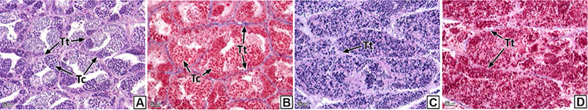
Similar to the ovaries, anatomical neoplasia was not confirmed in the testes. However, in histological analysis, neoplasia was observed in the form of groupings of basophilic sperm combined with connective tissues in the outside of the testicular outer membrane (Fig. 5). Testicular neoplasm was prevalent in 37.6% of the male samples (Table 2).

dr-25-3-123-g5
Fig. 5. Photomicrographs of testis in the rock bream, Oplegnathus fasciatus. (A) Normal testis. (B) Testicular neoplasm. H-E stain. Section showing the formation of testicular tissue and spermatids (Sd) in the outer testicular membrane (Tm).
Download Original Figure

The testis of teleosts consist of multiple testicular lobules surrounded by thin connective tissues (Lee & Huh, 2000; Blazer, 2002). Each testicular lobule retains numerous testicular cysts, and germ cells develop synchronously in the same testicular cyst (Lee et al., 2010). Spermatogenesis in teleosts is controlled by the central nervous system and endocrinological function of Leydig cells and Sertoli cells spread around the testis (Grier, 1981). Therefore, the deformation of testicular lobules and cysts in the rock bream could be determined to be histological evidence of an inability to obtain normal spermatogenesis.

4 Scale formation in the gonadal cavity

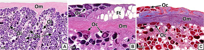
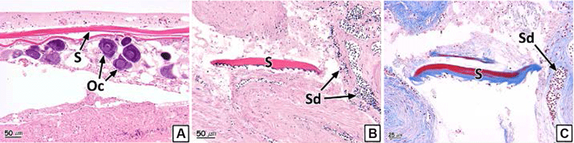
We also observed the formation of scales inside the gonadal cavity in some individual samples (Fig. 6). The scales showed eosinophilic stainability in the H-E stain and the center of the scales turned red and the peripheral parts stained to blue in Masson's trichrome stain (Fig. 6B and C). The prevalence of scale formation in the ovary and testis was 2.0% and 2.1% respectively (Table 2).

dr-25-3-123-g6
Fig. 6. Photomicrographs of scale (S) formation in the ovary (A) and testis (B) and (C) of the rock bream, Oplegnathus fasciatus. (A) and (B) H-E stain, (C) Masson's trichrome stain. Oc, oocytes; Sd, spermatids.
Download Original Figure

Scales of teleosts are formed by the hardening of connective tissues in the dermis of the skin (Takashima & Hibiya, 1995); moreover, vital somatic tissues inside the gonads also consist of connective tissues (Genten et al., 2009). Chemical factors such as EDC’s (endocrine disrupting chemicals) affects influence adipogenesis and osteogenesis in mesenchymal stem cells (Bateman et al., 2017). Chemical factors influence the synthesis of collagen, a major component of connective tissue, in fish such as Salmo gairdneri, Salvelinus fontinalis, Pimephales promrlas, Ictalurus punctatus (Mayer et al., 1977) and Pimephales promelas (Mehrle et al., 1981). In addition, in association with other gonad abnormalities identified in this study, one of the important causes of scale formation in the gonads is considered to be chemicals. Although the formation of scales inside the gonads in rock bream resulted from the disruption of the endocrine system due to chemical factors during the early embryonic stage, especially when connective tissues are differentiated, additional research regarding this topic is required.

5 Intersexuality

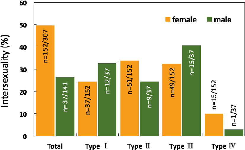
The overall intersexuality in the rock breams reached 42.2%, being higher in the female samples (49.5% in females and 26.2% in males; Fig. 7).

dr-25-3-123-g7
Fig. 7. Intersexuality of the rock bream, Oplegnathus fasciatus.
Download Original Figure

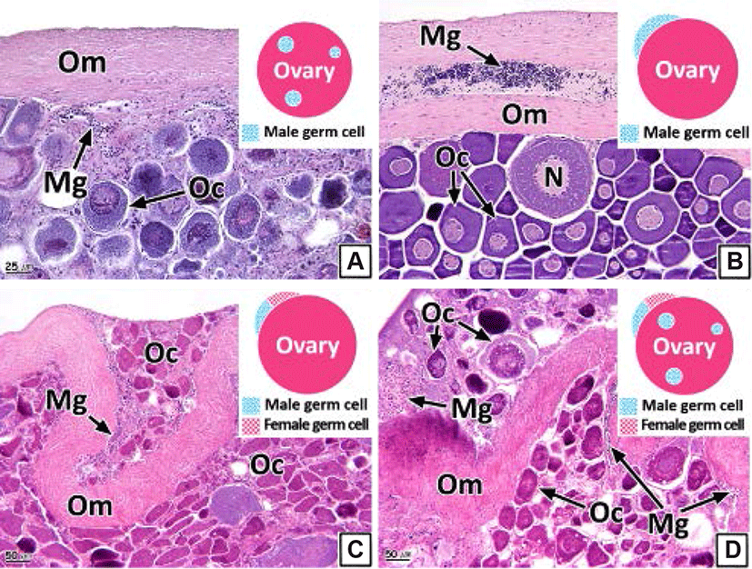
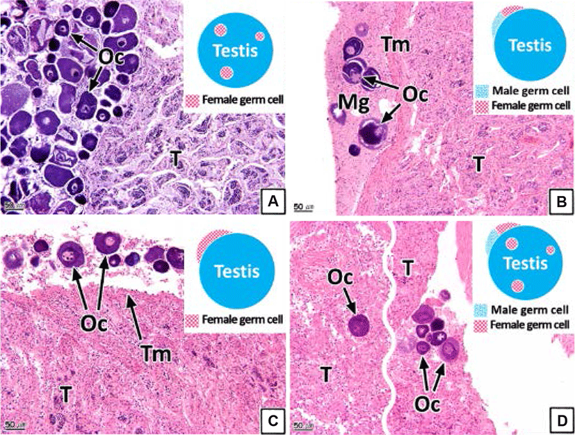
The intersex type observed in the gonads was histologically classified into four types (I, II, III, and IV). Type I shows germ cells of the opposite sex spread throughout the connective tissues of the gonadal cavity (Figs. 8A and 9A). Type II is the distribution of germ cells of the opposite sex in the newly formed connective tissues outside the gonad outer membrane (Figs. 8B and 9B). Type III represents the form in which the sperm and oocytes coexist in the connective tissues outside the gonad outer membrane (Figs. 8C and 9C). Type IV is the form in which the sperm and oocytes coexist in connective tissues outside the gonad outer membrane as well as in the gonadal cavity (Figs. 8D and 9D).

dr-25-3-123-g8
Fig. 8. Schematic diagram and photomicrographs of intersex type in the female rock bream, Oplegnathus fasciatus. (A) Type I. (B) Type II. (C) Type III. (D) Type IV. H-E stain. Mg, male germ cells; N, nucleus; Oc, oocytes; Om, ovarian outer membrane.
Download Original Figure
dr-25-3-123-g9
Fig. 9. Schematic diagram and photomicrographs of intersex type in the male rock bream, Oplegnathus fasciatus. (A) Type I. (B) Type II. (C) Type III. (D) Type IV. H-E stain. Mg, male germ cells; Oc, oocytes; T, testis; Tm, testicular outer membrane.
Download Original Figure

Intersex is one of the pathological sexual abnormalities that manifest due to environmental factors, such as the presence of endocrine-disrupting chemicals or heavy metals (Bortone & Davis, 1994). Studies of such abnormalities in teleosts have been undertaken in various species including Gambusia affinis and largemouth bass (Drysdale & Bortone, 1989; Blazer, 2002; Barnhoorn et al., 2010; Lee et al., 2010; Bahamonde et al., 2013; Mankidy et al., 2013).

Two primary causes of intersexuality and structural abnormalities in the gonads of rock bream can be confirmed from the results of the present study. The first cause is the presence of chemical factors such as EDC’s in their habitat. The second cause is the chemical components contained in mixed feeds, which are artificially supplied during the early life stages of artificial production of young rock bream. However, more detailed studies are required to ascertain the reasons for intersexuality and gonad structural abnormalities.

Acknowledgements

Not applicable.

Conflict of interests

The authors declare no potential conflict of interest.

Authors' contributions

Con ceptualization: Choi JS, Kim Y, Lee JS.

Data curation: Choi JS, Kim H.

Formal analysis: Shin SR, Kim HJ, Kim H.

Methodology: Lee JS.

Software: Shin SR, Kim HJ.

Validation: Kim Y, Lee JS.

Investigation: Choi JS, Shin SR, Kim HJ, Kim H.

Writing-original draft: Choi JS, Kim Y, Lee JS.

Writing-review & editing: Shin SR, Kim HJ, Lee JS.

Ethics approval

This article does not require IRB/IACUC approval because there are no human and animal participants.

REFERENCES

1.

Bahamonde PA, Munkittrick KR, Martyniuk CJ. 2013; Intersex in teleost fish: Are we distinguishing endocrine disruption from natural phenomena?. Gen Comp Endocrinol. 192:192-25

2.

Barber BJ. 1996; Effects of gonadal neoplasms on oogenesis in softshell clams, Mya arenaria.. J Invertebr Pathol. 67:161-168

3.

Barnhoorn IEJ, van Dyk JC, Pieterse GM, Bornman MS. 2010; Intersex in feral indigenous freshwater Oreochromis mossambicus, from various parts in the Luvuvhu River. Limpopo Province South Africa. Ecotoxicol Environ Saf. 73:1537-1542

4.

Bateman ME, Strong AL, McLachlan JA, Burow ME, Bunnell BA. 2017; The effects of endocrine disruptors on adipogenesis and osteogenesis in mesenchymal stem cells: A review.. Front Endocrinol. 7(171)

5.

Blazer VS. 2002; Histopathological assessment of gonadal tissue in wild fishes.. Fish Physiol Biochem. 26:85-101

6.

Bortone SA, Davis WP. 1994; Fish intersexuality as indicator of environmental stress.. Bioscience. 44:165-172

7.

Carballal MJ, Barber BJ, Iglesias D, Villalba A. 2015; Neoplastic diseases of marine bivalves.. J Invertebr Pathol. 131:83-106

8.

Colin N, Porte C, Fernandes D, Barata C, Padrós F, Carrassón M, Monroy M, Cano-Rocabayera O, de Sostoa A, Piña B, Maceda-Veiga A. 2016; Ecological relevance of biomarkers in monitoring studies of macro-invertebrates and fish in Mediterranean rivers.. Sci Total Environ. 540:307-323

9.

Cuevas N, Zorita I, Costa PM, Quincoces I, Larreta J, Franco J. 2015; Histopathological indices in sole (Solea solea) and hake (Merluccius merluccius) for implementation of the European Marine Strategy Framework Directive along the Basque continental shelf (SE Bay of Biscay).. Mar Pollut Bull. 94:185-198

10.

Drysdale DT, Bortone SA. 1989; Laboratory induction of intersexuality in the mosquitofish, Gambusia affinis. using paper mill effluent. Bull Environ Contam Toxicol. 43:611-617

11.

FIRA (Korea Fisheries Resources Agency) (2015) Release discharge of Gyeongsangnam-do, Jeollannam-do of the rock bream. Oplegnathus fasciatus in 2012–2015. .

12.

Fregeneda-Grandes JM, García-Iglesias MJ, Pérez-Martínez C, Aller-Gancedo JM. 2010; Mixed germ cell-stromal testicular neoplasia in a crucian carp (Carassius carassius L.): A case report.. Bull Eur Assoc Fish Pathol. 30:114-119.

13.

Genten F, Terwinghe E, Danguy A. 2009; Atlas of Fish Histology.. Science, Enfield, NH,. pp:1-215.

14.

Grier HJ. 1981; Cellular organization of the testis and spermatogenesis in fishes.. Am Zool. 21:345-357

15.

Huggett RJ, Kimerle RA, Mehrle PM, Bergman HL. 1992; Biomarkers: Biochemical, Physiological and Histological Markers of Anthropogenic Stress.. Lewis, Boca Raton, FL,. pp:1-347.

16.

Kim IS, Choi Y, Lee CL, Lee YJ, Kim BJ, Kim JH. 2005; Illustrated Book of Korean Fishes.. Kyo-Hak, Seoul, Korea. pp:1-613.

17.

Kokokiris L, Ragias V, Athanassopoulou F. 2005; Histopathological alterations of gonads in teleosts.. J Hell Vet Med Soc. 56:138-153

18.

Lee JS, Huh SH. 2000; Reproductive biology of the slimy, Leiognathus nuchalis (Teleostei: Leiognathidae).. Korean J Ichthyol. 12:192-202.

19.

Lee JS, Kim JW, Park JJ, Ju SM, Park JS, Lee DG, Yun TW, Choi KH, Yoon JH, Eom IC. 2010; Sex ratio and intersexuality in coastal fishes near industrial complex of Korea.. J Fish Pathol. 23:211-219.

20.

Mankidy R, Wiseman S, Ma H, Giesy JP. 2013; Biological impact of phthalates.. Toxicol Lett. 217:50-58

21.

Mayer FL, Mehrle PM, Schoettger RA. Collagen metabolism in fish exposed to organic chemicals.. In In: Tubb RA, editor. 1977; Recent Advances in Fish Toxicology: A Symposium. U.S. Environmental Protection Agency. CorvallisOR pp:31-54.

22.

Mehrle PM, Mayer FL, Buckler DR. 1981; Kepone and mirex: Effects on bone development and swim bladder composition in fathead minnows.. Trans Am Fish Soc. 110:638-643

23.

National Research Council (1983) Risk Assessment in the Federal Government: Managing the Process. National Academies PressWashington DC pp:1-191.

24.

Pinkney AE, Myers MS, Rutter MA. 2017; Histopathology of brown bullhead (Ameiurus nebulosus), smallmouth bass (Micropterus dolomieu), and yellow perch (Perca flavescens) in relation to polychlorinated biphenyl (PCB) contamination in the Hudson River.. Sci Total Environ. 575:1325-1338

25.

Schmitt CJ, Hinck JE, Blazer VS, Denslow ND, Dethloff GM, Bartish TM, Coyle JJ, Tillitt DE. 2005; Environmental contaminants and biomarker responses in fish from the Rio Grande and its U.S. tributaries: Spatial and temporal trends.. Sci Total Environ. 350:161-193

26.

Smolarz K, Hallmann A, Zabrzańska S, Pietrasik A. 2017; Elevated gonadal atresia as biomarker of endocrine disruptors: Field and experimental studies using Mytilus trossulus (L.) and 17-alpha ethinylestradiol (EE2).. Mar Pollut Bull. 120:58-67

27.

Spitsbergen JM, Buhler DR, Peterson TS. 2012; Neoplasia and neoplasm: Associated lesions in laboratory colonies of zebrafish emphasizing key influences of diet and aquaculture system design.. ILAR J. 53:114-125

28.

Takashima F, Hibiya T. 1995; An Atlas of Fish Histology: Normal and Pathological Features..2nd ed.Kodansha. Tokyo, Japan: pp p. 1-195.