Development & Reproduction
Korean Society of Developmental Biology
Short communication

Lin28 and Imp are Required for Stability of Bowl Transcripts in Hub Cells of the Drosophila Testis

Van To1https://orcid.org/0000-0002-3296-4344, Hyun Ju Kim1https://orcid.org/0000-0003-4819-7431, Wijeong Jang1https://orcid.org/0000-0002-3056-4566, Perinthottathil Sreejith1https://orcid.org/0000-0001-5445-3265, Changsoo Kim1,https://orcid.org/0000-0002-2852-9649
1School of Biological Sciences and Technology, Chonnam National University, Gwangju 61186, Korea
Corresponding author Changsoo Kim, School of Biological Sciences and Technology, Chonnam National University, Gwangju 61186, Korea. Tel: +82-62-530-5201, E-mail: changgk2001@hanmail.net

© Copyright 2021 The Korean Society of Developmental Biology. This is an Open-Access article distributed under the terms of the Creative Commons Attribution Non-Commercial License (http://creative-commons.org/licenses/by-nc/3.0/) which permits unrestricted non-commercial use, distribution, and reproduction in any medium, provided the original work is properly cited.

Received: Oct 15, 2021 ; Revised: Nov 28, 2021 ; Accepted: Dec 15, 2021

Published Online: Dec 31, 2021

Abstract

Hub cells comprise a niche for germline stem cells and cyst stem cells in the Drosophila testis. Hub cells arise from common somatic gonadal precursors in embryos, but the mechanism of their specification is still poorly understood. Here we find that RNA binding proteins Lin28 and Imp mediate transcript stability of Bowl, a known hub specification factor; Bowl transcripts were reduced in the testis of Lin28 and Imp mutants, and also when RNA-mediated interference against Lin28 or Imp was expressed in hub cells. In tissue culture Luciferase assays involving the Bowl 3’UTR, stability of Luc reporter transcripts depended on the Bowl 3’UTR and required Lin28 and Imp. Our findings suggest that proper Bowl function during hub cell specification requires Lin28 and Imp in the testis hub cells.

Keywords: Lin28; Imp; Niche; Stem cells; Testis; Bowl; Drosophila

INTRODUCTION

Adult stem cells are present in most tissues and provide differentiated cells when needed throughout an animal’s entire life (Kretzschmar & Clevers, 2017). This ability is realized through a stem cell’s unique capability for self-renewing cell division, in which it divides to simultaneously generate a daughter stem cell and a daughter cell that initiates differentiation (Funk et al., 2020). This unique property of stem cells is provided by the niche, a local environment that houses stem cells: a daughter cell within the niche’s area of influence maintains stemness and does not undergo differentiation, while one outside the niche loses stemness and starts differentiation (Spradling et al., 2008; Losick et al., 2011). Accordingly, stem cells are attached to niche cells and influenced by signaling molecules from them.

The Drosophila testis offers an incisive genetic animal model for dissection of molecular mechanisms underlying niche specification, aging, and niche-stem cell interaction (Herrera & Bach, 2019). An assembly of ~12 cells known as hub cells is located at the terminal tip of the testis and provides the niche for both germline stem cells (GSCs) and cyst stem cells (CySCs) (Yamashita et al., 2005). Hub cells are directly attached to GSCs and CySCs via adherens junctions, and express and secrete signaling molecules that stimulate GSCs and CySCs to promote stemness, cell division, and their attachment to hub cells (Kiger et al., 2001; Tulina & Matunis, 2001; Shivdasani & Ingham, 2003; Kawase et al., 2004; Leatherman & DiNardo, 2010; Michel et al., 2011).

Within the Drosophila testis, the RNA binding protein Lin28 is exclusively expressed in hub cells (Sreejith et al., 2019). Lin28 not only acts as a repressor of Let-7 biogenesis but also controls the stability and translation of target mRNAs including Unpaired (Upd), an extrinsic stem cell self-renewal factor that is expressed and secreted from hub cells (Kiger et al., 2001; Tulina & Matunis, 2001; Sreejith et al., 2019). A second RNA binding protein from a conserved protein family, IGF-II messenger RNA binding protein (Imp), is also required for stabilizing Upd transcripts (Toledano et al., 2012). However, what other factors Lin28 and Imp may act to stabilize in hub cells is yet largely unknown.

Hub cells and CySCs are both specified during embryogenesis from somatic gonadal precursors (SGPs) (D Le Bras & Van Doren, 2006; Kitadate & Kobayashi, 2010; iNardo et al., 2011; Okegbe & DiNardo, 2011). For hub cells, specification, aggregation, and assembly are known to require Notch signaling and the transcription factor Bowl, while CySC specification requires Lines (DiNardo et al., 2011; Okegbe & DiNardo, 2011). Notably, the embryonic gonads of Bowl mutants contain fewer hub cells, while increased Bowl activity transforms CySCs to the hub cell fate (DiNardo et al., 2011); these findings indicate that Bowl is a hub cell specification factor. Similarly, Lin28 mutant embryonic gonads contain fewer hub cells (Sreejith et al., 2019), suggesting that Lin28 could function in hub cell specification together with Bowl. Here we find that in hub cells, Bowl transcripts require Lin28 and Imp for stability.

RESULTS AND DISCUSSION

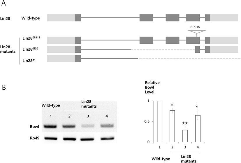
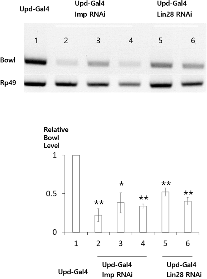
Lin28 was previously shown to be required for stabilizing transcripts of Upd, an important hub cell factor (Sreejith et al., 2019). This observation prompted us to identify other targets that are stabilized by Lin28 in hub cells of the Drosophila testis. To identify new target transcripts, we carried out reverse transcription polymerase chain reaction (RT-PCR) and compared transcript levels in Lin28 mutant and wild-type testis. This screening identified Bowl transcripts as reduced in the Lin28 mutant (Fig. 1). Lin28 is exclusively expressed in hub cells, suggesting that Bowl reduction is due to Lin28 mutation specifically in hub cells. To confirm this notion with an independent method, we used the bipartite GAL4-UAS system (Brand et al., 1994) in combination with RNA-mediated interference (RNAi) to reduce Lin28 function exclusively in hub cells. Transgenic flies bearing Upd-Gal4 (hub cell-specific Gal4) and UAS-Lin28 RNAi were generated (Upd-Gal4>UAS-Lin28 RNAi), in which Lin28 RNAi was specifically expressed in hub cells. RT-PCR of testis samples from two independent Lin28 RNAi lines confirmed the reduction of Bowl transcripts (Fig. 2). We likewise examined the relation of Bowl and Imp; however, Imp null mutation was lethal, so we instead relied on Imp knockdown in hub cells of the adult testis. Testis from three independent lines of Upd-Gal4>UAS-Imp RNAi flies expressing Imp RNAi in hub cells likewise showed reduction of Bowl transcripts (Fig. 2). Taken together, these data suggest that maintenance of Bowl transcripts in hub cells requires both Lin28 and Imp.

dr-25-4-313-g1
Fig. 1. Reduced Bowl transcripts in Lin28 mutant testis. (A) Lin28 mutants with a P-element insertion in the 4th exon (Lin28EP915) and with deletions in the middle of an exon as indicated (Lin28df30 and Lin28∆1). (B) (Left) Agarose gel image of RT-PCR products from testis with Lin28 mutations. Rp49 used as a loading control. 1. Wild type, 2. Lin28EP915, 3. Lin28df30, 4. Lin28∆1. (Right) Quantification of RT-PCR bands by Image J analysis, with Bowl band intensity normalized by control Rp49 band intensity. 1. Wild type, 2. Lin28EP915, 3. Lin28df30, 4. Lin28∆1. p-values by student’s t-test in Sigma-Aldrich Plot. * p<0.05, ** p<0.01. RT-PCR, reverse transcription polymerase chain reaction.
Download Original Figure
dr-25-4-313-g2
Fig. 2. Reduced Bowl transcripts in the testis with Lin28 and Imp RNAi expressed in the hub cells. (Up) Agarose gel image of RT-PCR products from testis with RNAi expressed in hub cells. Rp49 used as a loading control. 1. Upd-Gal4, 2–4. Upd-Gal4>UAS-Imp RNAi (2032, 20322, 34977), 5–6. Upd-Gal4>UAS-Lin28 RNAi (50679, 29564). (Down) Quantification of RT-PCR bands by Image J analysis, with Bowl band intensity normalized by control Rp49 band intensity. 1. Upd-Gal4, 2–4. Upd-Gal4>UAS-Imp RNAi (2032, 20322, 34977), 5–6. Upd-Gal4>UAS-Lin28 RNAi (50679, 29564). p-values by student’s t-test in Sigma-Aldrich Plot. * p<0.05, ** p<0.01. RT-PCR, reverse transcription polymerase chain reaction; RNAi, RNA-mediated interference; Upd, unpaired.
Download Original Figure

To further investigate the molecular mechanisms underlying promotion of Bowl transcript stability by Lin28 and Imp, we carried out cell culture experiments using Luciferase (Luc) constructs bearing the Bowl 3’UTR (Luc-Bowl 3’UTR), employing RT-PCR to examine whether those hybrid transcripts were stabilized by Lin28 and Imp, which stabilizes target mRNAs through 3’UTR (Toledano et al., 2012; Lee et al., 2019; Sreejith et al., 2019). When either Lin28 or Imp was co-transfected with the construct, Luc-Bowl 3’UTR transcript levels were increased (Fig. 3A). Meanwhile, levels of control Luc transcripts lacking the Bowl 3’UTR were not affected by the presence of either Lin28 or Imp (Fig. 3B). These results suggest that Lin28 and Imp stabilize Luc-Bowl 3’UTR transcripts through the Bowl 3’UTR.

dr-25-4-313-g3
Fig. 3. Bowl 3’UTR mediates stability of Bowl transcripts. Agarose gel image of RT-PCR products from S2 cells co-transfected with the Luc-Bowl 3’UTR hybrid construct (A) and parental Luc construct (B) in combination with parental vector (1), Lin28 expression vector (2), Imp expression vector (3) and Lin28+Imp (4). Rp49 used as a loading control. Primers used for RT-PCR indicated as arrows. (Bottom) Quantification of RT-PCR bands by Image J analysis, with Bowl band intensity normalized by control Rp49 band intensity. p-values by student’s t-test in Sigma-Aldrich Plot. * p<0.05, ** p<0.01. RT-PCR, reverse transcription polymerase chain reaction.
Download Original Figure

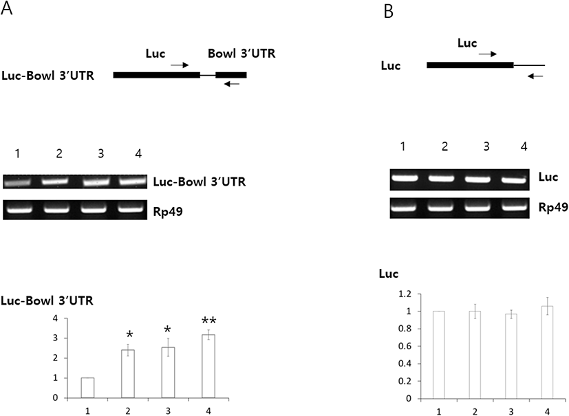
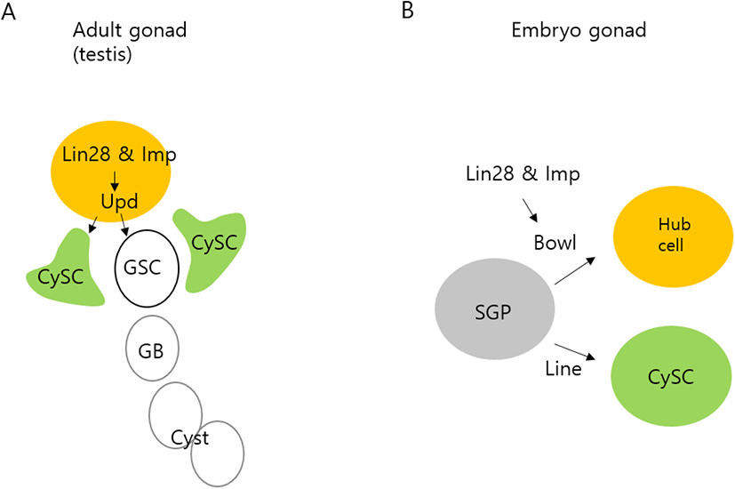
Fig. 4 illustrates our proposed model of the roles of Lin28 and Imp in the testis. We and others previously showed that in adult testis hub cells, Lin28 and Imp are required to sustain hub cell maintenance and function and act through stabilizing mRNAs encoding Upd, a critical hub cell factor that stimulates stemness and cell division of GSCs and CySCs (Fig. 4A) (Toledano et al., 2012; Sreejith et al., 2019). In this communication, we identify a new hub cell-specific mRNA stabilized by Lin 28 and Imp, the Bowl transcript (Fig. 4B). Bowl was previously shown to be a hub cell specification factor during early development of the embryonic gonad (DiNardo et al., 2011). Our findings suggest that Lin28 and Imp are present in hub cells and act to protect both Bowl transcripts in embryos and Upd transcripts in adults. An interesting question remains regarding the mechanistic particulars of how Lin28 and Imp stabilize Upd and Bowl mRNAs. Future studies are required to address this question.

dr-25-4-313-g4
Fig. 4. Models depicting roles of Lin28 and Imp in the Drosophila testis. (A) Roles of Lin28 and Imp in hub cells of the adult testis. Upd transcripts are stabilized by Lin28 and Imp. GSC, germline stem cell; CySC, cyst stem cell; GB, goniablast. (B) Roles of Lin28 and Imp in hub cell specification in the embryonic gonads. Bowl transcripts are stabilized by Lin28 and Imp. SGP, somatic gonadal precursor. Upd, unpaired.
Download Original Figure

MATERIALS AND METHODS

1. Drosophila strains

Flies were grown in standard fly food. W1118, Lin28EP915, and ImpMI05901 were from the Bloomington Drosophila Stock Center (BDSC). Lin28df30 was from F. Michon and Lin28∆1 from N. Sokol. UAS-Lin28 RNAi (50679, 29564) and UAS-Imp RNAi (34977) were from the BDSC and UAS-Imp RNAi (20321, 20322) from the Vienna Drosophila RNAi Center (VDRC).

2. Reverse transcription polymerase chain reaction (RT-PCR)

RNA was extracted using standard TRIzol (Invitrogen) procedure. Complementary DNA synthesis was performed with oligodT primers using one microgram RNA. PCR was performed with the following primers: Rp49: Forward 5’-CACCAGGAACTTCTTGAATCCGG-3’, Re v erse 5’-A GA T C G T GAA GAA GC GC A C C-3’; Bo wl: F or war d 5’-CTGCTCATCCACGAGAGG-3’, Reverse 5’-TGTGGACAGCCAAGGTTC-3’; Luc-Bowl 3’UTR: Forward 5’-CC CTCGAG CAATCCATTAATGGAG-3’, Reverse 5’-GC TCTAGA CATTATTAATGCATACTTTATTTGA-3’. RT-PCR were carried out three replicates in Fig. 1, 2, and 3.

3. Cloning of Luc-Bowl 3’UTR

pAc5.1A-Luc (S2 cell expression vector) was created using the Luc gene from the pGL3-Basic vector (Promega, Madison, WI, USA), which was ligated into the EcoRI-NotI sites of pAc5.1/V5-HisA (Invitrogen). The Bowl 3’UTR (853 bp) from Bowl cDNA (RE05342, Drosophila Genetic Resource Centre) was ligated into the XhoI-XbaI sites of the pAc5.1A-Luc vector.

4. S2 cell culture and transfection

Schneider 2 cells were grown and maintained in Shields and Sang M3 insect Media (Sigma-Aldrich, St. Louis, MO, USA) supplemented with 10% insect media supplement (Sigma-Aldrich) with appropriate antibiotics at 25 °C. Cells were subcultured every five days at a 1:5 ratio when confluency reached around 100%. The DDAB method of transfection was employed with some modifications for 24-well plates (Han, 1996). Cells were collected and assayed three days after transfection.

Conflict of interests

The authors declare no potential conflict of interest.

Acknowledgements

This work was supported by grants from the National Research Foundation of Korea (NRF-2021R1A2C1010334 to CK, NRF-2020R1I1A1A01074292 to WJ, NRF-2017R1A6A3A11032327 to WJ). We thank the Bloomington Drosophila Stock Center (BDSC) and Vienna Drosophila RNAi Center (VDRC) for flies, and the Drosophila Genetic Resource Center (DGRC) for Bowl cDNA. We are grateful to F. Michon for sharing Lin28df30, T. Guy for Upd-Gal4, and N Sokol for Lin28∆1.

Authors’ contributions

Conceptualization: Kim C.

Data curation: To V, Kim HJ.

Methodology: Jang W, Sreejith P.

Writing-original draft: To V, Kim HJ

Writing-review & editing: Kim C.

Ethics approval

This article does not require IRB/IACUC approval because there are no human and animal participants.

REFERENCES

1.

Brand AH, Manoukian AS, Perrimon N. 1994; Ectopic expression in Drosophila. Methods Cell Biol. 44:635-654

2.

DiNardo S, Okegbe T, Wingert L, Freilich S, Terry N. 2011; Lines and bowl affect the specification of cyst stem cells and niche cells in the Drosophila testis. Development. 138:1687-1696

3.

Funk MC, Zhou J, Boutros M. 2020; Ageing, metabolism and the intestine. EMBO Rep. 21:e50047

4.

Han K. 1996; An efficient DDAB-mediated transfection of Drosophila S2 cells. Nucleic Acids Res. 24:4362-4363

5.

Herrera SC, Bach EA. 2019; JAK/STAT signaling in stem cells and regeneration: From Drosophila to vertebrates. Development. 146:dev167643

6.

Kawase E, Wong MD, Ding BC, Xie T. 2004; Gbb/Bmp signaling is essential for maintaining germline stem cells and for repressing bam transcription in the Drosophila testis. Development. 131:1365-1375

7.

Kiger AA, Jones DL, Schulz C, Rogers MB, Fuller MT. 2001; Stem cell self-renewal specified by JAK-STAT activation in response to a support cell cue. Science. 294:2542-2545

8.

Kitadate Y, Kobayashi S. 2010; Notch and Egfr signaling act antagonistically to regulate germ-line stem cell niche formation in Drosophila male embryonic gonads. Proc Natl Acad Sci USA. 107:14241-14246

9.

Kretzschmar K, Clevers H. 2017; Wnt/β-catenin signaling in adult mammalian epithelial stem cells. Dev Biol. 428:273-282

10.

Le Bras S, Van Doren M. 2006; Development of the male germline stem cell niche in Drosophila. Dev Biol. 294:92-103

11.

Leatherman JL, DiNardo S. 2010; Germline self-renewal requires cyst stem cells and stat regulates niche adhesion in Drosophila testes. Nat Cell Biol. 12:806-811

12.

Lee M, Nguyen TMT, Kim K. 2019; In-depth study of lin-28 suggests selectively conserved let-7 independent mechanism in Drosophila. Gene. 687:64-72

13.

Losick VP, Morris LX, Fox DT, Spradling A. 2011; Drosophila stem cell niches: A decade of discovery suggests a unified view of stem cell regulation. Dev Cell. 21:159-171

14.

Michel M, Raabe I, Kupinski AP, Pérez-Palencia R, Bökel C. 2011; Local BMP receptor activation at adherens junctions in the Drosophila germline stem cell niche. Nat Commun. 2:415

15.

Okegbe TC, DiNardo S. 2011; The endoderm specifies the mesodermal niche for the germline in Drosophila via Delta-Notch signaling. Development. 138:1259-1267

16.

Shivdasani AA, Ingham PW. 2003; Regulation of stem cell maintenance and transit amplifying cell proliferation by tgf-β signaling in Drosophila spermatogenesis. Curr Biol. 13:2065-2072

17.

Spradling AC, Nystul T, Lighthouse D, Morris L, Fox D, Cox R, Tootle T, Frederick R, Skora A. 2008; Stem cells and their niches: Integrated units that maintain Drosophila tissues. Cold Spring Harb Symp Quant Biol. 73:49-57

18.

Sreejith P, Jang W, To V, Hun Jo Y, Biteau B, Kim C. 2019; Lin28 is a critical factor in the function and aging of Drosophila testis stem cell niche. Aging. 11:855-873

19.

Toledano H, D’Alterio C, Czech B, Levine E, Jones DL. 2012; The let 7-Imp axis regulates ageing of the Drosophila testis stem-cell niche. Nature. 485:605-610

20.

Tulina N, Matunis E. 2001; Control of stem cell self-renewal in Drosophila spermatogenesis by JAK-STAT signaling. Science. 294:2546-2549

21.

Yamashita YM, Fuller MT, Jones DL. 2005; Signaling in stem cell niches: Lessons from the Drosophila germline. J Cell Sci. 118:665-672