Research article

Potent Influence of Exogenous Melatonin on In Vitro Oocyte Maturation in the Longchin Goby, Chaenogobius annularis

Dae Guen Kim1,*https://orcid.org/0000-0001-8064-4612, In Joon Hwang2,*https://orcid.org/0000-0001-5740-3925, Hea Ja Baek3,https://orcid.org/0000-0002-4578-5919
Author Information & Copyright
1Gyeongsangnamdo Fisheries Resources Research Institute, Tongyeong 50411, Korea
2West Sea Fisheries Research Institute, National Institute of Fisheries Science, Incheon 22383, Korea
3Department of Marine Biology, Pukyong National University, Busan 48513, Korea

* These authors contributed equally to this work.

Corresponding author Hea Ja Baek, Department of Marine Biology, Pukyong, National University, Busan 48513, Korea, Tel: +82-51-629-5924, Fax: +82-51-629-5931, E-mail: hjbaek@pknu.ac.kr

© Copyright 2023 The Korean Society of Developmental Biology. This is an Open-Access article distributed under the terms of the Creative Commons Attribution Non-Commercial License (http://creative-commons.org/licenses/by-nc/3.0/) which permits unrestricted non-commercial use, distribution, and reproduction in any medium, provided the original work is properly cited.

Received: Jul 07, 2023 ; Revised: Aug 23, 2023 ; Accepted: Sep 11, 2023

Published Online: Sep 30, 2023

Abstract

Effects of changes in photoperiod on the reproductive events in fish are suggested to be mediated mainly via the action of melatonin (MEL). Changing levels of plasma MEL throughout the day and year are suggested to influence the hypothalamus–pituitary–gonadal axis in fish. Therefore, in this study, we aimed to investigate the effects of MEL on oocyte maturation and germinal vesicle breakdown (GVBD) in the marine fish, Chaenogobius annularis, in vitro. Oocytes at three different stages (pre-, mid-, and late-vitellogenesis) were incubated with (a) only MEL (5, 10, 50, 100, 500, and 1,000 pg/mL) and (b) 50 pg/mL of 17α,20β-dihydroxy-4-pregnen-3-one (17α20βP), maturation-inducing hormone (MIH) of this species, and MEL (4-h incubation before addition of MIH). Any single MEL treatment did not significantly induce GVBD. However, treatment with 50 pg/mL MEL or MIH significantly induced GVBD. These results suggest that preincubation with MEL accelerates the effect of MIH on longchin goby oocyte maturation.

Keywords: Melatonin; Oocyte; Maturation; Longchin goby

INTRODUCTION

Melatonin (N-acetyl-5-methoxytryptamine, MEL), a neuroendocrine hormone synthesized and secreted from the pineal gland of the brain, plays a significant physiological role in the regulation of fish gonadal development (Tosini & Menaker, 1996; Borjigin et al., 1999; Mondal et al., 2017). In vertebrates, including teleosts, plasma MEL, and photoperiod-dependent neurohormones in fish (Goldman, 2001; Reiter et al., 2009), levels are rhythmically expressed through a 24 h light-dark cycle, as the plasma MEL concentration is low during the daytime and high during the nighttime (Falcón et al., 1987; Bolliet et al., 1996; Migaud et al., 2010; Ogiwara & Takahashi, 2016).

MEL mediates the relationship between circadian rhythms and the hypothalamic-pituitary-gonadal (H-P-G) axis, thereby regulating reproductive events, including oocyte development and maturation (Vera et al., 2006; Falcón et al., 2007, 2010; Tamura et al., 2014). In other words, the pineal gland translates environmental signals, such as the photoperiod, into rhythmic changes in hormonal fluctuations in the H-P-G axis; photoreceptor cells of the pineal gland produce MEL in a day-length-dependent manner (Falcón et al., 2007; Brüning et al., 2018). MEL receptors are distributed in all brain areas, including the pituitary gland, retina, and many other tissues, such as the ovary (Falcón et al., 2010; Hong et al., 2014). These studies suggested that MEL exerts direct effects on teleost ovaries by activating MEL receptors. MEL acts directly at the ovarian level to modulate ovarian physiology, including steroidogenesis (Adriaens et al., 2006; Wang et al., 2019), folliculogenesis (Choi et al., 2015), oocyte maturation (Maitra et al., 2013) and ovulation (Ogiwara & Takahashi, 2016). In medaka ovaries, Ogiwara & Takahashi (2016) reported that pre-ovulatory follicles are capable of synthesizing MEL. They found that MEL is constitutively synthesized in the granulosa cells of preovulatory follicles during the 24 h spawning cycle of fish (Takahashi & Ogiwara, 2021). Moreover, MEL has been detected in fish ovaries (Hasan et al., 2014).

Therefore, the direct effects of MEL on the gonads of fish were supported. Chattoraj et al. (2005, 2008) showed that pre-incubation with MEL accelerated the action of maturation inducing hormone (MIH) during oocyte maturation; 17α,20β-dihydroxy-4-pregnen-3-one (17α20βP) has been considered to be MIH in most teleost fish (Patiño & Sullvian, 2002; Nagahama & Yamashita, 2008). Similarly, the influence of MEL on the process of oocyte maturation has been reported in zebrafish; exposure of ovarian follicles to MEL significantly increased the rate of germinal vesicle breakdown (GVBD) in oocytes of MIH-treated follicles (Carnevali et al., 2011). Incubation of oocytes with MEL diminishes the stimulatory effect of MEL and MIH on the rate of GVBD (Maitra & Hasan, 2016). However, the specific mechanisms by which MEL directly regulates reproductive events remain unclear.

Therefore, we examined the direct effects of exogenous MEL on longchin goby oocytes at different stages of maturation (pre-, mid-, and late-vitellogenesis) by investigating the GVBD rate in vitro. In addition, we examined whether preincubation with MEL accelerates MIH-induced maturation. These trials were performed based on the previously reported ranges of circulating MEL levels (Vera et al., 2006; Nikaido et al., 2009; Oliveira et al., 2011).

MATERIALS AND METHODS

1. Experimental fish

Experimental fish (body length: 5.1–6.5 cm) were captured from tide pool at Cheong-sa-po, Busan, Korea, during reproductive period (March to May).

2. Incubation protocols for the determination of germinal vesicle breakdown (GVBD)

Ovaries were dissected from several mature females, rinsed with ice-cold balanced salt solution (132.96 mM NaCl, 3.09 mM KCl, 0.28 mM MgSO4 · 7H2O, 0.98 mM MgCl2 · 6H2O, 3.40 mM CaCl2 · 6H2O, and 3.65 mM HEPES), and separated into different maturation stages according to the diameters measuring with an ocular micrometer under a dissecting microscope. The extracted ovaries were categorized into three different maturation stages: pre-vitellogenesis (0.61–0.66 mm of oocyte diameter), mid-vitellogenesis (0.71–0.74 mm of oocyte diameter), and late-vitellogenesis (0.80–0.86 mm of oocyte diameter) (Baek et al., 2007).

Oocytes were incubated in each well of 24-well culture plates containing 1 mL of Leibowitz L-15 medium (Gibco, Grand Island, NY, USA) with (a) MEL at the concentrations of 5, 10, 50, 100, 500, and 1,000 pg/mL, (b) 50 ng/mL of 17α20βP which is MIH of this species, or (c) both 50 ng/mL of 17α20βP and MEL (4 h pre-incubated before addition of 17α20βP, at the concentration 50, 100, and 500 pg/mL). The pH and osmolarity of the media were adjusted to 7.7 and 300 mOsm, respectively. Each plate was cultured for different incubation times of 24 h (mid- and late-vitellogenesis) or 48 h (pre-vitellogenesis) at 18°C with constant gentle shaking. Each treatment was conducted in three wells with 20 oocytes per well and all experiments were repeated three times.

At the end of the incubation period, oocytes were fixed with a clearing solution (ethanol: formalin: glacial acetic acid=6:3:1). The effects of the treatments on oocyte maturation were determined by GVBD using a dissecting microscope (BX-50, Olympus, Tokyo, Japan).

3. Statistical analysis

GVBD results are expressed as the means with the SEM and examined using the Kruskal– Wallis test followed by Bonferroni adjustment. Statistical significance was set at p<0.05.

RESULTS

1. Morphological observation of oocytes before in vitro incubation

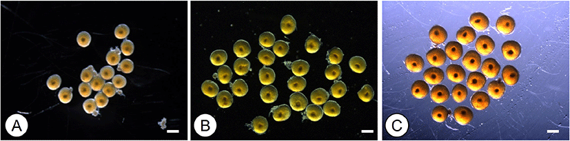
Oocytes isolated from the ovaries of the longchin goby are shown in Fig. 1. Each developmental stage of oocytes was observed under a microscope immediately before incubation. In pre- and mid-vitellogenic oocytes, GVs were located in the middle of the cytoplasm (Fig. 1A and B). GVs of late-vitellogenic oocytes were still in the middle of the cytoplasm, and migrating GVs were not observed (Fig. 1C).

dr-27-3-127-g1
Fig. 1. Morphology of longchin goby oocytes before in vitro incubation. Germinal vesicles (GVs) were seen near the center of oocytes. A, Pre-vitellogenesis; B, mid-vitellogenesis; C, late-vitellogenesis. Scale bar indicates 500 μm.
Download Original Figure
2. Effects of melatonin (MEL) on germinal vesicle breakdown (GVBD)

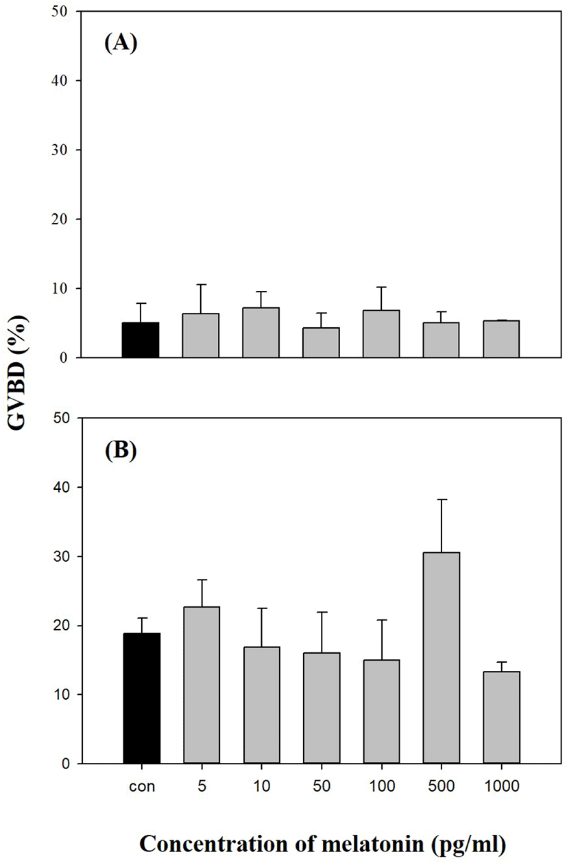
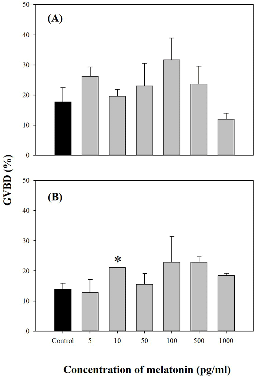
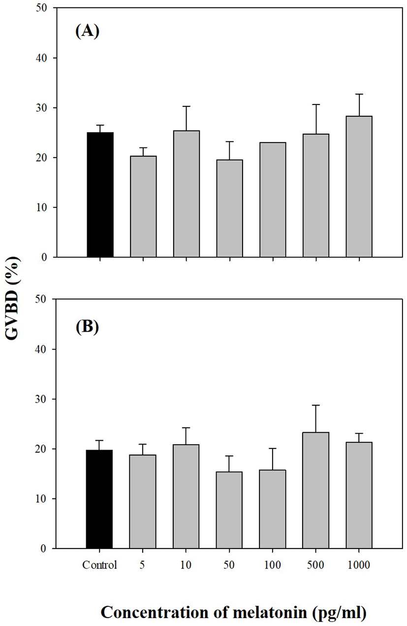
We investigated the direct effects of exogenous MEL on longchin goby oocytes at various stages of maturation. As shown in Figs. 24, MEL did not show any significant effect on GVBD, except for one experiment in 0.74 mm diameter oocytes were cultured with 10 pg/mL MEL (p<0.05).

dr-27-3-127-g2
Fig. 2. Effects of different melatonin concentrations on in vitro germinal vesicle breakdown (GVBD) in pre-vitellogenic oocytes of longchin goby after 48 h of incubation. Values are represented as the mean±SE of each GVBD in three replicate wells with 20 oocytes/well. A, oocyte diameter of 0.61 mm; B, oocyte diameter of 0.66 mm.
Download Original Figure
dr-27-3-127-g3
Fig. 3. Effects of different melatonin concentrations on in vitro GVBD in mid-vitellogenic oocytes of longchin goby after 24 h of incubation. Values are represented as the mean±SE of each GVBD in three replicate wells with 20 oocytes/well. Data were analyzed using the Kruskal–Wallis and Tukey tests. Asterisks indicate the significant differences from controls (p<0.05). A, oocyte diameter of 0.71 mm; B, oocyte diameter of 0.74 mm. GVBD, germinal vesicle breakdown.
Download Original Figure
dr-27-3-127-g4
Fig. 4. Effects of different melatonin concentrations on in vitro GVBD in late-vitellogenic oocytes of longchin goby after 24 h of incubation. Values are represented as the mean±SE of each GVBD in three replicate wells with 20 oocytes/well. A, oocyte diameter of 0.80 mm; B, oocyte diameter of 0.86 mm. GVBD, germinal vesicle breakdown.
Download Original Figure
3. Effects melatonin (MEL) with maturation inducing hormone (MIH) on germinal vesicle breakdown (GVBD)

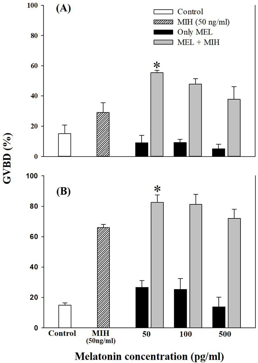
Oocytes of late-vitellogenesis stage (0.80 and 0.86 mm) were incubated with 50, 100, and 500 pg/mL MEL alone or in combination with 50 ng/mL MIH (17α20βP; Fig. 5). Significant GVBD induction was observed with 50 ng/mL MEL combined with 50 ng/mL MIH (p<0.05). Other higher levels of MEL treatment (both 100 and 500 pg/mL) with 50 ng/mL MIH showed an increase in GVBD compared to single treatment with 50 ng/mL MIH, although the difference was not significant.

dr-27-3-127-g5
Fig. 5. Effects of different melatonin concentrations and maturation-inducing hormone (MIH; 50 ng/mL) on in vitro GVBD in late-vitellogenic oocytes of longchin goby after 24 h of incubation. Values are represented as the mean±SE of each GVBD in three replicate wells with 20 oocytes/well. Data were analyzed using the Kruskal–Wallis and Tukey tests. Asterisks indicate the significant differences from MIH (50 ng/mL; p<0.05). A, oocyte diameter of 0.80 mm; B, oocyte diameter of 0.86 mm. MIH, maturation inducing hormone; GVBD, germinal vesicle breakdown.
Download Original Figure

DISCUSSION

A recent review by Takahashi & Ogiwara (2021) showed that MEL, a neurohormone primarily synthesized and secreted in the pineal glands of vertebrates following a circadian rhythm, is an important regulator of various physiological processes, including reproduction. Moreover, in teleosts, this hormone is synthesized in the ovary and acts directly at the ovary level to regulate ovarian physiology, including steroid hormone synthesis, oocyte maturation, and ovulation (Maitra & Hasan, 2016; Mondal et al., 2017).

In teleosts, MEL has been detected in the ovaries of Indian carp (Calta catla) (Hasan et al., 2014; Mondal et al., 2017), medaka (Oryzias latipes) (Ogiwara & Takahashi, 2016), and zebrafish (Danio rerio) (Yumnamcha et al., 2017; Khan et al., 2018). In these studies, serum MEL levels were low during the day and high at night. MEL circulating levels range from 146.6 to 697.7 pg/mL in seabream (Diplodus puntazzo) (Vera et al., 2006), 69 to 224 pg/mL in Mozambique tilapia (Oreochromis mossambicus) (Nikaido et al., 2009), and 34.1 to 287.6 pg/mL in Senegal sole (Solea senegalensis) (Oliveira et al., 2011), even though reared conditions are different, under various light and temperature conditions. Based on these levels, our experiments set concentrations in the range of 5–1,000 pg/mL in vitro. Based on the in vitro study of Chattoraj et al. (2005), the pre-incubation time of MEL was set to 4 h. Incubation time of goby oocytes was set to 24–48 h according to our previous studies (Baek et al., 2007; Baek, 2008).

In this experiment, the direct treatments of MEL alone at concentrations of 5, 10, 50, 100, 500, and 1,000 pg/mL in vitro using the oocytes of pre-vitellogenesis (0.61–0.66 mm diameter), mid-vitellogenesis (0.71 and 0.74 mm diameter), and late-vitellogenesis (0.81 and 0.86 mm diameter) did not evoke any response to GVBD except 0.74 mm diameter oocytes cultured with 10 pg/mL MEL (p<0.05). This could be due to differences in the developmental stages of the oocytes. We suspect that the oocytes of 0.74 mm were more sensitive to the action of MEL than the oocytes of other sizes, and the extent of the stimulatory effect on GVBD depends on MEL. However, the combination of MEL and MIH in late-vitellogenesis (0.81 and 0.86 mm) oocytes increased the rate of GVBD compared to other experimental groups (treated with MIH or MEL alone); 50 pg/mL MEL had maximum acceleratory effect on MIH induced GVBD of oocytes. These results indicated that the activity of MIH-induced GVBD was accelerated after 4 h of pre-incubation with MEL. Similar results regarding the direct effects of MEL on fish oocyte maturation, including GVBD, have been reported by Chattoraj et al. (2005), who showed that pre-incubation with MEL accelerated the action of MIH during oocyte maturation in carp (Labeo rohita and Cyprinus carpio). Similarly, during oocyte maturation in zebrafish, the exposure of ovarian follicles to MEL significantly increased the GVBD rate of MIH-treated follicles (Carnevali et al., 2011). Conversely, the incubation of oocytes with MEL diminished the stimulatory effect of MEL and MIH on the rate of GVBD in Catla catla (Chattoraj et al., 2008; reviewed by Maitra & Hasan, 2016).

Exogenous MEL treatment leads to elevated intraovarian MEL titers, which may stimulate endogenously available antioxidants to reduce the oxidative stress in the carp ovary and accelerate (during the preparatory phase) or retard (during the pre-spawning or spawning phase) the growth of oocytes (Mondal et al., 2017). Takahashi & Ogiwara (2021) reported that the direct action of MEL affects ovarian steroidogenesis in some teleosts; for example, using a mudskipper with fully grown ovarian follicle during the spawning season, plasma 17α20βP levels were significantly increased after MEL injection (Hong et al., 2014). They also showed that MEL directly stimulated ovarian fragments in vitro to produce significantly higher progestin levels. Takahashi & Ogiwara (2021) suggested that MEL affects oocyte maturation via its antioxidant activity. However, the molecular mechanisms underlying the direct effects of MEL remain largely unknown.

Carps treated with MEL at various phases of the reproductive cycle showed correlations among ovarian MEL, different antioxidants, and different stages of oocyte growth (Mondal et al., 2017). Further research is needed to prove the roles of MEL, both in its direct action on the ovary and its indirect effects via the H-P-G axis, in fish reproduction.

Conflict of interests

The authors declare no potential conflict of interest.

Acknowledgements

This study was supported by a Research Grant of Pukyong National University (2021).

Authors’ contributions

Conceptualization: Kim DG, Baek HJ.

Data curation: Kim DG, Hwang IJ.

Formal analysis: Kim DG, Hwang IJ.

Methodology: Kim DG.

Software: Hwang IJ.

Validation: Hwang IJ.

Investigation: Kim DG, Baek HJ.

Writing-original draft: Kim DG, Hwang IJ, Baek HJ.

Writing-review & editing: Kim DG, Hwang IJ, Baek HJ.

Ethics approval

The animal study was reviewed and approved by Animal Ethics Committee of Pukyong National University (PKNU; Regulation No. 554).

REFERENCES

1.

Adriaens I, Jacquet P, Cortvrindt R, Janssen K, Smitz J. 2006; Melatonin has dose-dependent effects on folliculogenesis, oocyte maturation capacity and steroidogenesis. Toxicology. 228:333-343

2.

Baek H. 2008; Production of C21-steroids in longchin goby, Chasmichthys dolichognathus during oocyte maturation. Dev Reprod. 12:107-112.

3.

Baek HJ, Hwang IJ, Kim KS, Lee YD, Kim HB, Yoo MS. 2007; Effects of BPA and DES on longchin goby (Chasmichthys dolichognathus) in vitro during the oocyte maturation. Mar Environ Res. 64:79-86

4.

Bolliet V, Ali MA, Lapointe FJ, Falcón J. 1996; Rhythmic melatonin secretion in different teleost species: An in vitro study. J Comp Physiol B. 165:677-683

5.

Borjigin J, Li X, Snyder SH. 1999; The pineal gland and melatonin: Molecular and pharmacologic regulation. Annu Rev Pharmacol Toxicol. 39:53-65

6.

Brüning A, Kloas W, Preuer T, Hölker F. 2018; Influence of artificially induced light pollution on the hormone system of two common fish species, perch and roach, in a rural habitat. Conserv Physiol. 6:coy016

7.

Carnevali O, Gioacchini G, Maradonna F, Olivotto I, Migliarini B. 2011; Melatonin induces follicle maturation in Danio rerio. PLoS One. 6:e19978

8.

Chattoraj A, Bhattacharyya S, Basu D, Bhattacharya S, Bhattacharya S, Maitra SK. 2005; Melatonin accelerates maturation inducing hormone (MIH): Induced oocyte maturation in carps. Gen Comp Endocrinol. 140:145-155

9.

Chattoraj A, Seth M, Maitra SK. 2008; Influence of serotonin on the action of melatonin in MIH-induced meiotic resumption in the oocytes of carp Catla catla. Comp Biochem Physiol A Mol Integr Physiol. 150:301-306

10.

Choi YJ, Kim BS, Choi CY. 2015; Effect of long-after glow phosphorescent pigment on repro-ductive parameters and ovarian maturation in the yellowtail damselfish, Chrysiptera parasema. Comp Biochem Physiol A Mol Integr Physiol. 182:113-120

11.

Falcón J, Besseau L, Sauzet S, Boeuf G. 2007; Melatonin effects on the hypothalamo–pituitary axis in fish. Trends Endocrinol Metab. 18:81-88

12.

Falcón J, Guerlotté JF, Voisin P, Collin JPH. 1987; Rhythmic melatonin biosynthesis in a photoreceptive pineal organ: A study in the pike. Neuroendocrinology. 45:479-486

13.

Falcón J, Migaud H, Muñoz-Cueto JA, Carrillo M. 2010; Current knowledge on the melatonin system in teleost fish. Gen Comp Endocrinol. 165:469-482

14.

Goldman BD. 2001; Mammalian photoperiodic system: Formal properties and neuroendocrine mechanisms of photoperiodic time measurement. J Biol Rhythms. 16:283-301

15.

Hasan KN, Moniruzzaman M, Maitra SK. 2014; Melatonin concentrations in relation to oxidative status and oocyte dynamics in the ovary during different reproductive phases of an annual cycle in carp Catla catla. Theriogenology. 82:1173-1185

16.

Hong LY, Hong WS, Zhu WB, Shi Q, You XX, Chen SX. 2014; Cloning and expression of melatonin receptors in the mudskipper Boleophthalmus pectinirostris: Their role in synchronizing its semilunar spawning rhythm. Gen Comp Endocrinol. 195:138-150

17.

Khan ZA, Labala RK, Yumnamcha T, Devi SD, Mondal G, Devi HS, Rajiv C, Bharali R, Chattoraj A. 2018; Artificial light at night (ALAN), an alarm to ovarian physiology: A study of possible chronodisruption on zebrafish (Danio rerio). Sci Total Environ. 628–629:1407-1421

18.

Maitra SK, Chattoraj A, Mukherjee S, Moniruzzaman M. 2013; Melatonin: A potent candidate in the regulation of fish oocyte growth and maturation. Gen Comp Endocrinol. 181:215-222

19.

Maitra SK, Hasan KN. 2016; The role of melatonin as a hormone and an antioxidant in the control of fish reproduction. Front Endocrinol. 7:38

20.

Migaud H, Davie A, Taylor JF. 2010; Current knowledge on the photoneuroendocrine regulation of reproduction in temperate fish species. J Fish Biol. 76:27-68

21.

Mondal P, Hasan KN, Pal PK, Maitra SK. 2017; Influences of exogenous melatonin on the oocyte growth and oxidative status of ovary during different reproductive phases of an annual cycle in carp Catla catla. Theriogenology. 87:349-359

22.

Nagahama Y, Yamashita M. 2008; Regulation of oocyte maturation in fish. Dev Growth Differ. 50:S195-S219

23.

Nikaido Y, Ueda S, Takemura A. 2009; Photic and circadian regulation of melatonin production in the Mozambique tilapia Oreochromis mossambicus. Comp Biol Physiol A Mol Integr Physiol. 152:77-82

24.

Ogiwara K, Takahashi T. 2016; A dual role for melatonin in medaka ovulation: Ensuring prosta-glandin synthesis and actin cytoskeleton rearrangement in follicular cells. Biol Reprod. 94:64

25.

Oliveira C, Mañanós E, Ramos J, Sánchez-Vázquez FJ. 2011; Impact of photoperiod manipulation on day/night changes in melatonin, sex steroids and vitellogenin plasma levels and spawning rhythms in Senegal sole, Solea senegalensis. Comp Biochem Physiol A Mol Integr Physiol. 159:291-295

26.

Patiño R, Sullivan CV. 2002; Ovarian follicle growth, maturation, and ovulation in teleost fish. Fish Physiol Biochem. 26:57-70

27.

Reiter RJ, Tan DX, Manchester LC, Paredes SD, Mayo JC, Sainz RM. 2009; Melatonin and reproduction revisited. Biol Reprod. 81:445-456

28.

Takahashi T, Ogiwara K. 2021; Roles of melatonin in the teleost ovary: A review of the current status. Comp Biochem Physiol A Mol Integr Physiol. 254:110907

29.

Tamura H, Takasaki A, Taketani T, Tanabe M, Lee L, Tamura I, Maekawa R, Aasada H, Yamagata Y, Sugino N. 2014; Melatonin and female reproduction. J Obstet Gynaecol Res. 40:1-11

30.

Tosini G, Menaker M. 1996; Circadian rhythms in cultured mammalian retina. Science. 272:419-421

31.

Vera LM, Madrid JA, Sánchez-Vázquez FJ. 2006; Locomotor, feeding and melatonin daily rhythms in sharpsnout seabeam (Diplodus puntazzo). Physiol Behav. 88:167-172

32.

Wang X, Meng K, He Y, Wang H, Zhang Y, Quan F. 2019; Melatonin stimulates STAR expression and progesterone production via activation of the PI3K/AKT pathway in bovine theca cells. Int J Biol Sci. 15:404-415

33.

Yumnamcha T, Khan ZA, Rajiv C, Devi SD, Mondal G, Devi HS, Bharali R, Chattoraj A. 2017; Interaction of melatonin and gonadotropin-inhibitory hormone on the zebrafish brain-pituitary-reproductive axis. Mol Reprod Dev. 84:389-400