ARTICLE

Polo-Like Kinases (Plks), a Key Regulator of Cell Cycle and New Potential Target for Cancer Therapy

Su-Yeon Lee, Chuljoon Jang, Kyung-Ah Lee†
Author Information & Copyright
Corresponding author: Kyung-Ah Lee, Department of Biomedical Science, College of Life Science, CHA University, Seoul 135-081, Republic of Korea. Tel. : +82-2-557-3937, Fax : +82-2-563-2028, E-mail: leeka@ovary.co.kr

Copyright © 2014 © Copyright A Official Journal of the Korean Society of Developmental Biology. All Rights Reserved.. This is an Open Access article distributed under the terms of the Creative Commons Attribution Non-Commercial License (http://creativecommons.org/licenses/by-nc/3.0/) which permits unrestricted non-commercial use, distribution, and reproduction in any medium, provided the original work is properly cited.

Received: Jan 20, 2014 ; Revised: Feb 10, 2014 ; Accepted: Feb 15, 2014

ABSTRACT

Cell cycle process is regulated by a number of protein kinases and among them, serine/threonine kinases carry phosphate group from ATP to substrates. The most important three kinase families are Cyclin-dependent kinase (Cdk), Polo-like kinase (Plk), and Aurora kinase. Polo-like kinase family consists of 5 members (Plk1-Plk5) and they are involved in multiple functions in eukaryotic cell division. It regulates a variety of aspects such as, centrosome maturation, checkpoint recovery, spindle assembly, cytokinesis, apoptosis and many other features. Recently, it has been reported that Plks are related to tumor development and over-expressed in many kinds of tumor cells. When injected the anti-Plk antibody into human cells, the cells show aneuploidy, and if inhibit Plks, most of the mitotic cell division does not proceed properly. For that reasons, many inhibitors of Plk have been recently emerged as new target for remedy of the cancer therapeutic research. In this paper, we reviewed briefly the characteristics of Plk families and how Plks work in regulating cell cycles and cancer formation, and the possibilities of Plks as target for cancer therapy.


INTRODUCTION

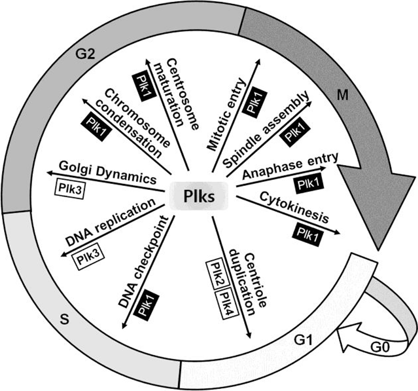
Plk is a serine/threonine kinase, which was firstly identified in Drosophila as Polo mutant over 27 years ago (Sunkel & Glover, 1988). This kinase is well conserved from budding yeast (Cdc5) to Drosophila (polo), Xenopus (Plxs) and mammals (Plks) (de Carcer et al., 2011a). In mammalian cells, it has been reported that four Plk family members, from Plk1 to Plk4, are expressed and recently Plk5 has been characterized in the mouse and human cells (de Carcer et al., 2011a). Plks are key regulators for various cellular events during cell division including centrosome maturation, DNA checkpoint activation, mitotic entry, spindle assembly, and cytokinesis (Fig. 1) (Glover et al., 1998; Dai, 2005; Strebhardt & Ullrich, 2006).
dr-18-1-65-g001
Fig. 1. Various cellular functions related with each Plk family member. Plk1, the first identified and the major Plk member, plays a role in mitotic entry, spindle assembly, anaphase entry and cytokinesis in mitotic phase and DNA checkpoint, chromosome condensation and centrosome maturation in interphase. In G1 phase, Plk2 and Plk4 promote centriole duplication and in S phase, Plk3 regulates DNA replication. Plk5, not mentioned above figure, is known as involved in neuron differentiation.
Download Original Figure

Plk1 has a crucial role in the regulation of mitotic checkpoints. Hence, overexpression of this kinase causes overriding checkpoints, which can lead to immature cell division with aneuploidy, a hallmark of cancer (Kops et al., 2005). Plk1 overexpression contributes to cancer development by promoting excessive cell proliferation through the misregulation of check point functions (Degenhardt & Lampkin, 2010). Furthermore, Plk2 and Plk3 also have a relationship with p53 and are activated by DNA damage in mitosis, whereas Plk1 is inhibited (Jang et al., 2007). Plk2 is a direct transcriptional target of p53 and Plk3-mediated phosphorylation of p53 at Ser-20 is reported to stabilize p53 (Xie et al., 2001; Burns et al., 2003). Therefore, Plk families might be a good target for cancer therapy and several Plk inhibitors are in clinical studies recently. In this article, we review characteristics and functions of Plk family members in regard with cell cycles, carcinogenesis, and apoptosis, as well as with the possibilities of Plks as cancer therapeutic targets.

Plk Family Members

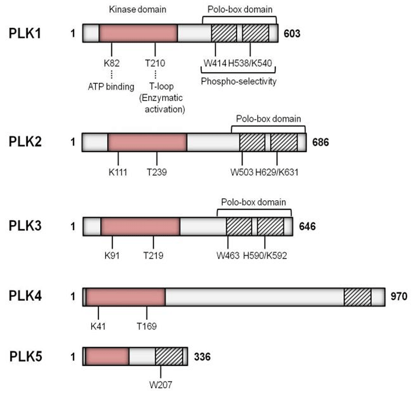
The Plks have a highly conserved N-terminal catalytic domain and C-terminal domain with one or more polo box domains (PBDs) (Fig. 2) (Strebhardt & Ullrich, 2006). In the yeast and Drosophila, only a single Plk gene has been identified to date, while the higher vertebrates have multiple Plk genes of which Plk1 is most similar to Drosophila polo (Ohkura et al., 1995). Plk2 (also known as SNK) localizes at the centrosomes and plays a role in the entry into S phase (de Carcer et al., 2011a). Inhibition of Plk2 resulted in formation of aberrant number of centrioles (Warnke et al., 2004). Plk3 (also named FNK or PRK) is required at the G1-S phase transition and for DNA replication (Zimmerman & Erikson, 2007). Plk4 (also known as SAK23) is found in all animals and it acts as a critical regulator of centriole duplication both in Drosophila and mammals (Kleylein-Sohn et al., 2007). Both the mouse and human Plk5 genes encode proteins, however, to some extent they are different that the mouse Plk5 contains a complete kinase domain and PBD, while the human Plk5 encloses only a small portion of the kinase domain along with one PBD (de Carcer et al., 2011b).
dr-18-1-65-g002
Fig. 2. Schematic diagram of the five Plk family genes in the human. The lengths of amino-acid are shown as numbers on the right. The kinase domains of proteins are described as pink box at N-terminus and the polo-box domains are shown at C-terminus as box with stripes. Key residues for ATP-binding and enzymatic activation (T-loop) within the kinase domains, and for phospho-selectivity within the polo-box domains are also indicated.
Download Original Figure
Plks in Cell Cycle Regulation

Mammalian Plks are required for the functional matu-ration of centrosomes and the assembly of a bipolar spindle. Microinjection of Plk1-specific antibody resulted in abnormal chromatin and microtubules, and hence in block of cell division (Lane & Nigg, 1996). These results were derived from small, unseparated centrosome. Shortly before the onset of mitosis, the size and number of centrosomes increase and they are required for organizing the microtubules (Karsenti et al., 1984). The functional maturation of centrosomes can be visualized by monitoring the accumulation of multiple proteins including γ-tubulin and M-phase-specific phosphoprotein (MPM-2) reactive phosphoepitopes (Lajoie-Mazenc et al., 1994). Plk1 anti-body-injected cells showed much smaller mitotic spindle poles and reduced expression of γ-tubulin and MPM-2 (Lane & Nigg, 1996).

Through recent studies, γ-tubulin is thought to be a critical element in centrosomal microtubule nucleation during late G2/early prophase (Shu & Joshi, 1995; Sunkel et al., 1995). Plk1 depletion or inhibition prevented the recruitment of γ-tubulin to centrosomes (Lane & Nigg, 1996; Haren et al., 2009). In addition, the accumulation of MPM-2 reactivity at centrosomes is inhibited in Plk1-injectied cells (Lane & Nigg, 1996).

In the previous study, it has been shown that the removal of centrosomes from mammalian cells in culture prevented every part of features of mitosis, including the activation of Cdc2 kinase (Maniotis & Schliwa, 1991). Cdc25C is an activator of Cdc2 kinase and dephosphorylates and activates the CyclinB-Cdc2 complex shortly before the entry into the mitosis. Both the DNA damage and DNA replication checkpoints regulate CyclinB-Cdc2 activation partly through Cdc25C. Plx1, Xenopus homolog of Plk, can phosphorylate and activate Cdc25C in vitro. In Xenopus oocyte maturation, Plx1 is activated in vivo with the same kinetics as Cdc25C (Qian et al., 1998). Depletion of Plx1 activity in Xenopus oocytes significantly inhibited the activation of Cdc25C and CyclinB-Cdc2 and inhibition of Plx1 in early Xenopus embryos resulted in an arrest of cleavage with abnormal spindles (Qian et al., 1998). These studies demonstrated that Plx1 plays important roles both in the activation of Cdc25C in the mitosis as well as in meiosis.

Through in vitro study in mammalian cells and Drosophila system, it has been known that Plk1 is required for important steps in the onset and completion of mitosis, such as bipolar spindle assembly, segregation of chromosomal DNA (Ohkura et al., 1995), centrosome maturation (Lane & Nigg, 1996) and anaphase promoting complex regulation (Descombes & Nigg, 1998). Recently, nuclear distribution gene C (NudC) and centrosomal ninein-like protein (Nlp) have been identified as Plk1 target proteins (Zhou et al., 2003). Plk1 phosphorylates NudC and regulates cytokinesis (Zhou et al., 2003). Nlp, a substrate of Plk1, interacts with the γ-tubulin ring complex and stimulates microtubule nucleation (Casenghi et al., 2003).

Plk2 is expressed in mainly G1 phase and important for the reproduction of centrosomes and centriole duplication (Cizmecioglu et al., 2008; Cizmecioglu et al., 2012). Mutation in the Plk2 polo-box inhibits normal localization of centrosome and centriole duplication (Cizmecioglu et al., 2008). Interestingly, Plk2 and Plk4 have a functional interaction and Plk2 mediated centriole duplication is associated with a Plk4 activity (Cizmecioglu et al., 2008). In mouse, depletion of Plk2 impairs embryonic growth and normal placental development resulted from decreased cell proliferation (Ma et al., 2003).

Plk3 is also a key regulator of cell cycles during mitosis. In S phase, Plk3 regulates DNA replication with phosphorylation of related proteins, topoisomerase IIα and DNA polymerase δ (Iida et al., 2008). In addition, Plk3 regulates Golgi dynamics during G2 phase for mitotic entry in mitotic cells (Lopez-Sanchez et al., 2009).

Plks in Cancer Development

It has been well known that Plk1 expression increased in various cancer tissues such as non small cell lung cancer (Wolf et al., 1997), squamous cell carcinoma of the head and neck (Knecht et al., 1999), esophageal cancer, gastric cancer (Tokumitsu et al., 1999) and colorectal cancer (Macmillan et al., 2001). In Plk1 siRNA-treated HeLa cells, the cell proliferation was dramatically inhibited whereas the cell viability decreased (Liu & Erikson, 2003). Especially, apoptosis was induced in Plk1-depleted cancer cells (Liu & Erikson, 2003). These results demonstrate that Plk1 is related to the causes of cancer such as abnormal centrosome formation with abnormal cell cycles and apoptosis.

It is well known that centrosomes are critical in causing genetic instability in cancer cells (Brinkley, 2001). They cause the abnormalities in spindles and disturb the chromosome segregation, which are found with reflective alterations in key cellular functions, such as regulation of apoptosis, control of cell cycles, and regulation of cell growth. Centrosomal abnormalities have been detected in early forms of the human prostate cancer (Pihan et al., 2001). Extra centrosomes in cancer cells may lead to chromosome mis-arrangements and damages which are causes of aneuploidy. This condition may induce the loss of tumor suppressor genes or activate oncogenes (Spankuch-Schmitt et al., 2002). Therefore those authors suggested that the centrosomes are driving force for cancer formation, not a consequence of it.

Plk1 is also associates with apoptosis. Fas-associated death domain-containing protein (FADD) was identified as a first adapter molecule that bridges the Fas-receptor during apoptosis (Peter et al., 1999). The phosphorylation of FADD serves as a critical marker for the human cancer diagnosis (Shimada et al., 2005). Plk1 phosphorylates FADD at Ser-194 in response to taxol treatment and is regulated through a negative feedback loop by its substrate, FADD (Jang et al., 2011). In addition, Plk1 binds to the major tumor suppressor gene, p53, in cultured mammalian cells, and pro-apoptotic function of p53 is vanished via interaction with Plk1 (Ando et al., 2004). Inhibition of Plk1 function prevented centrosome maturation and induced cell death in carcinoma cells and dominant negative Plk1 induced tumor-selective apoptosis in tumor cell lines (Lane & Nigg, 1996). Overexpression of Plk3 suppressed cell proliferation and induced apoptosis (Conn et al., 2000).

Plks in Cancer Therapy

The precise role of Plk1 in cancer cell is not apparent yet. However, based on the above findings, Plk1 has a critical role in the development of cancer. Therefore, regulation of Plk1 function might be an important appliance in cancer therapy. Because Plk1 activates Cyclin B/Cdc2 complex via Cdc25C and stimulates cell proliferation, the silencing of Plk1 will result in an inactivation of Cyclin B/Cdc2 complex-mediated mitotic arrest followed by apoptosis. In Plk1 siRNA-treated cells, cell proliferation was reduced and apoptosis was increased (Liu & Erikson, 2003). Therefore, pharmacological inhibitors of Plk1 may be valuable tools for treatment of cancer (Degenhardt & Lampkin, 2010; McInnes & Wyatt, 2011).

Concluding Remarks

The fact that centrosome abnormalities in mitotic cells may lead to chromosome missorting and damage causing cancer suggests the possibility that Plk1, a key regulator of centrosome maturation, might regulate carcinogenesis and can be used as a therapeutic target for cancer treatment. In many clinical researches, Plk1 expression was enhanced in the human cancer cells, but detailed mechanism of Plk1 in cancer development is not clearly understood yet. Nowadays, Plk1 is emerging as an attractive target for anticancer agents. Furthermore, the collected information about the functions of the other Plk families suggests that we can now selectively inhibit the Plk1 activity without affecting other isoforms. Therefore, the more scrutinized research on each Plk family member and its regulation in normal mitotic as well as in meiotic cells may enlarge our knowledge in cell biology and also provide us an opportunity to find out the breakthrough for cancer treatment.

ACKNOWLEDGEMENT

This work was supported by the National Research Foundation of Korea (NRF) grant funded by the Korea government (MSIP) (2012-046741).

REFERENCES

1.

Ando K, Ozaki T, Yamamoto H, Furuya K, Hosoda M, Hayashi S, Fukuzawa M, Nakagawara A. Polo-like kinase 1 (Plk1) inhibits p53 function by physical interaction and phosphorylation. J Biol Chem. 2004; 279:25549-25561.

2.

Brinkley BR. Managing the centrosome numbers game: from chaos to stability in cancer cell division. Trends Cell Biol. 2001; 11:18-21.

3.

Burns TF, Fei P, Scata KA, Dicker DT, El-Deiry WS. Silencing of the novel p53 target gene Snk/Plk2 leads to mitotic catastrophe in paclitaxel (taxol)-exposed cells. Mol Cell Biol. 2003; 23:5556-5571.

4.

Casenghi M, Meraldi P, Weinhart U, Duncan PI, Korner R, Nigg EA. Polo-like kinase 1 regulates Nlp, a centrosome protein involved in microtubule nucleation. Dev Cell. 2003; 5:113-125.

5.

Cizmecioglu O, Krause A, Bahtz R, Ehret L, Malek N, Hoffmann I. Plk2 regulates centriole duplication through phosphorylation-mediated degradation of Fbxw7 (human Cdc4). J Cell Sci. 2012; 125:981-992.

6.

Cizmecioglu O, Warnke S, Arnold M, Duensing S, Hoffmann I. Plk2 regulated centriole duplication is dependent on its localization to the centrioles and a functional polo-box domain. Cell Cycle. 2008; 7:3548-3555.

7.

Conn CW, Hennigan RF, Dai W, Sanchez Y, Stambrook PJ. Incomplete cytokinesis and induction of apoptosis by overexpression of the mammalian polo-like kinase, Plk3. Cancer Res. 2000; 60:6826-6831.

8.

Dai W. Polo-like kinases, an introduction. Oncogene. 2005; 24:214-216.

9.

de Carcer G, Manning G, Malumbres M. From Plk1 to Plk5: functional evolution of polo-like kinases. Cell Cycle. 2011a; 10:2255-2262.

10.

de Carcer G, Escobar B, Higuero AM, Garcia L, Anson A, Perez G, Mollejo M, Manning G, Melendez B, Abad-Rodriguez J, Malumbres M. Plk5, a polo box domain-only protein with specific roles in neuron differentiation and glioblastoma suppression. Mol Cell Biol. 2011b; 31:1225-1239.

11.

Degenhardt Y, Lampkin T. Targeting Polo-like kinase in cancer therapy. Clin Cancer Res. 2010; 16:384-389.

12.

Descombes P, Nigg EA. The polo-like kinase Plx1 is required for M phase exit and destruction of mitotic regulators in Xenopus egg extracts. EMBO J. 1998; 17:1328-1335.

13.

Glover DM, Hagan IM, Tavares AA. Polo-like kinases: a team that plays throughout mitosis. Genes Dev. 1998; 12:3777-3787.

14.

Haren L, Stearns T, Luders J. Plk1-dependent recruitment of gamma-tubulin complexes to mitotic centrosomes involves multiple PCM components. PLoS One. 2009; 4:e5976.

15.

Iida M, Matsuda M, Komatani H. Plk3 phosphorylates topoisomerase II alpha at Thr(1342), a site that is not recognized by Plk1. Biochem J. 2008; 411:27-32.

16.

Jang MS, Lee SJ, Kim CJ, Lee CW, Kim E. Phosphorylation by polo-like kinase 1 induces the tumor-suppressing activity of FADD. Oncogene. 2011; 30:471-481.

17.

Jang YJ, Ji JH, Choi YC, Ryu CJ, Ko SY. Regulation of Polo-like kinase 1 by DNA damage in mitosis. Inhibition of mitotic PLK-1 by protein phosphatase 2A. J Biol Chem. 2007; 282:2473-2482.

18.

Karsenti E, Kobayashi S, Mitchison T, Kirschner M. Role of the centrosome in organizing the interphase microtubule array: properties of cytoplasts containing or lacking centrosomes. J Cell Biol. 1984; 98:1763-1776.

19.

Kleylein-Sohn J, Westendorf J, Le Clech M, Habedanck R, Stierhof YD, Nigg EA. Plk4-induced centriole biogenesis in human cells. Dev Cell. 2007; 13:190-202.

20.

Knecht R, Elez R, Oechler M, Solbach C, von Ilberg C, Strebhardt K. Prognostic significance of polo-like kinase (PLK) expression in squamous cell carcinomas of the head and neck. Cancer Res. 1999; 59:2794-2797.

21.

Kops GJ, Weaver BA, Cleveland DW. On the road to cancer: aneuploidy and the mitotic checkpoint. Nat Rev Cancer. 2005; 5:773-785.

22.

Lajoie-Mazenc I, Tollon Y, Detraves C, Julian M, Moisand A, Gueth-Hallonet C, Debec A, Salles-Passador I, Puget A, Mazarguil H, et al. Recruitment of antigenic gamma-tubulin during mitosis in animal cells: presence of gamma-tubulin in the mitotic spindle. J Cell Sci. 1994; 107(Pt 10):2825-2837.

23.

Lane HA, Nigg EA. Antibody microinjection reveals an essential role for human polo-like kinase 1 (Plk1) in the functional maturation of mitotic centrosomes. J Cell Biol. 1996; 135:1701-1713.

24.

Liu X, Erikson RL. Polo-like kinase (Plk)1 depletion induces apoptosis in cancer cells. Proc Natl Acad Sci U S A. 2003; 100:5789-5794.

25.

Lopez-Sanchez I, Sanz-Garcia M, Lazo PA. Plk3 interacts with and specifically phosphorylates VRK1 in Ser342, a downstream target in a pathway that induces Golgi fragmentation. Mol Cell Biol. 2009; 29:1189-1201.

26.

Ma S, Charron J, Erikson RL. Role of Plk2 (Snk) in mouse development and cell proliferation. Mol Cell Biol. 2003; 23:6936-6943.

27.

Macmillan JC, Hudson JW, Bull S, Dennis JW, Swallow CJ. Comparative expression of the mitotic regulators SAK and PLK in colorectal cancer. Ann Surg Oncol. 2001; 8:729-740.

28.

Maniotis A, Schliwa M. Microsurgical removal of centrosomes blocks cell reproduction and centriole generation in BSC-1 cells. Cell. 1991; 67:495-504.

29.

McInnes C, Wyatt MD. PLK1 as an oncology target: current status and future potential. Drug Discov Today. 2011; 16:619-625.

30.

Ohkura H, Hagan IM, Glover DM. The conserved Schizosaccharomyces pombe kinase plo1, required to form a bipolar spindle, the actin ring, and septum, can drive septum formation in G1 and G2 cells. Genes Dev. 1995; 9:1059-1073.

31.

Peter ME, Scaffidi C, Medema JP, Kischkel F, Krammer PH. The death receptors. Results Probl Cell Differ. 1999; 23:25-63.

32.

Pihan GA, Purohit A, Wallace J, Malhotra R, Liotta L, Doxsey SJ. Centrosome defects can account for cellular and genetic changes that characterize prostate cancer progression. Cancer Res. 2001; 61:2212-2219.

33.

Qian YW, Erikson E, Li C, Maller JL. Activated polo-like kinase Plx1 is required at multiple points during mitosis in Xenopus laevis. Mol Cell Biol. 1998; 18:4262-4271.

34.

Shimada K, Matsuyoshi S, Nakamura M, Ishida E, Konishi N. Phosphorylation status of Fas-associated death domain-containing protein (FADD) is associated with prostate cancer progression. J Pathol. 2005; 206:423-432.

35.

Shu HB, Joshi HC. Gamma-tubulin can both nucleate microtubule assembly and self-assemble into novel tubular structures in mammalian cells. J Cell Biol. 1995; 130:1137-1147.

36.

Spankuch-Schmitt B, Bereiter-Hahn J, Kaufmann M, Strebhardt K. Effect of RNA silencing of polo-like kinase-1 (PLK1) on apoptosis and spindle formation in human cancer cells. J Natl Cancer Inst. 2002; 94:1863-1877.

37.

Strebhardt K, Ullrich A. Targeting polo-like kinase 1 for cancer therapy. Nat Rev Cancer. 2006; 6:321-330.

38.

Sunkel CE, Gomes R, Sampaio P, Perdigao J, Gonzalez C. Gamma-tubulin is required for the structure and function of the microtubule organizing centre in Drosophila neuroblasts. EMBO J. 1995; 14:28-36.

39.

Tokumitsu Y, Mori M, Tanaka S, Akazawa K, Nakano S, Niho Y. Prognostic significance of polo-like kinase expression in esophageal carcinoma. Int J Oncol. 1999; 15:687-692.

40.

Warnke S, Kemmler S, Hames RS, Tsai HL, Hoffmann-Rohrer U, Fry AM, Hoffmann I. Polo-like kinase-2 is required for centriole duplication in mammalian cells. Curr Biol. 2004; 14:1200-1207.

41.

Wolf G, Elez R, Doermer A, Holtrich U, Ackermann H, Stutte HJ, Altmannsberger HM, Rubsamen-Waigmann H, Strebhardt K. Prognostic significance of polo-like kinase (PLK) expression in non-small cell lung cancer. Oncogene. 1997; 14:543-549.

42.

Xie S, Wu H, Wang Q, Cogswell JP, Husain I, Conn C, Stambrook P, Jhanwar-Uniyal M, Dai W. Plk3 functionally links DNA damage to cell cycle arrest and apoptosis at least in part via the p53 pathway. J Biol Chem. 2001; 276:43305-43312.

43.

Zhou T, Aumais JP, Liu X, Yu-Lee LY, Erikson RL. A role for Plk1 phosphorylation of NudC in cytokinesis. Dev Cell. 2003; 5:127-138.

44.

Zimmerman WC, Erikson RL. Finding Plk3. Cell Cycle. 2007; 6:1314-1318.