


INTRODUCTION
Tissue immune homeostasis depends on continuous interactions between immune cells and their surrounding microenvironment. The bone marrow (BM) is highly responsive to systemic changes in the host, which can influence hematopoietic stem/progenitor populations and shift the balance between myeloid and lymphoid cell production. Hyperglycemic conditions increase hematopoietic stem/progenitor and myeloid-derived suppressor cell (MDSC) populations in BM, whereas aging-associated changes in stromal interleukins (IL)-6 and transforming growth factor (TGF)-β signaling contribute to altered hematopoietic lineage output and reduced lymphopoiesis (Kim et al., 2019; Valletta et al., 2020). Alterations established during hematopoiesis may persist beyond the BM and influence the properties of immune cells that subsequently enter peripheral tissues. Within peripheral tissues, monocyte-derived macrophages can undergo further phenotypic and functional adaptation in response to local environmental signals, as demonstrated in the lung, liver, and peritoneal cavity (Scott et al., 2016; Mould et al., 2017; Louwe et al., 2021). Together these findings indicate that immune-cell composition and function can be influenced by both alterations established during hematopoiesis and additional cues encountered within peripheral tissues. Given the central role of local microenvironments in coordinating immune responses, understanding the factors that regulate these environments is important for defining how immune homeostasis is maintained or disrupted.
Glyoxalase 1 (GLO1) is an enzyme involved in cellular adaptation to metabolic and oxidative stress and in the maintenance of cellular homeostasis. Alterations in GLO1 expression and activity have been reported in a range of chronic pathological conditions associated with metabolic dysfunction and inflammation. In diabetes-associated tissue injury, reduced GLO1 expression has been observed in diabetic wounds, while restoration of GLO1 activity or expression improves the function of BM-derived progenitor cells and macrophages and facilitates tissue repair (Li et al., 2019; Jiang et al., 2022). GLO1 expression is also reduced in macrophage activated by LPS and IFN-γ, suggesting that changes in GLO1 are closely associated with inflammation conditions (Prantner et al., 2021). In diabetic ischemic tissue, GLO1 overexpression has been shown to restore macrophage infiltration and inflammatory responses while improving angiogenesis and blood-flow recovery, whereas GLO1 overexpression attenuates microglial inflammatory responses in a model of neuroinflammation (Liu et al., 2025). However, most previous studies have focused on the functional consequences of manipulating GLO1 within specific cell types or on pathological responses within individual tissues. The impact of GLO1 deficiency on the broader immune environment, particularly across hematopoietic and peripheral tissue compartments, remains largely unexplored.
In this study, we used Glo1 knockout (Glo1-KO) mice to examine the effects of GLO1 deficiency on BM hematopoietic composition, the pulmonary inflammation environment. Our findings reveal alterations in BM myeloid populations together with changes in the pulmonary inflammatory profile. These findings highlight GLO1 as a potential regulator of immune homeostasis, with a role in coordinating hematopoietic and peripheral immune environments.
MATERIALS AND METHODS
Female wild-type C57BL/6J mice (8–10 weeks old) were purchased from DooYeol Biotech (Seoul, Korea). Glo1-KO mice on a C57BL/6 background were generated by intercrossing heterozygous (Glo1+/−) mice obtained from the Korea Research Institute of Bioscience and Biotechnology (KRIBB, Daejeon, Korea). Genotyping was confirmed by PCR using tail genomic DNA with the following primers: Glo1 common 5′-CAACCTGGAGCTTTGGTGTGTTTG-3′, Glo1 wild 5′-GCAAACGTGAAGGCCTGAGTCTG-3′, Glo1 mutant 5′-CCACAACGGGTTCTTCTGTTAGTCC-3′. Mice were maintained under specific pathogen-free conditions with a 12-h light/dark cycle and ad libitum access to food and water at the Kangwon National University Laboratory Animal Center. All animal procedures were approved by the Institutional Animal Care and Use Committee of Kangwon National University (IACUC No. KW-221027-1).
BM cells were harvested from femurs by flushing with ice-cold RPMI-1640. The cell suspension was centrifuged at 800×g for 5 min, and the cell pellet was treated with RBC lysis buffer for 3 min. After washing with PBS containing 1% FBS, the cells were subjected to flow cytometric analysis.
For hematopoietic stem/progenitor cell analysis, BM cells were stained with V450 Mouse Lineage Antibody Cocktail (561301, BD Bioscience, Franklin Lakes, NJ, USA), anti-mouse Ly-6A/E(Sca-1) (558162, BD Bioscience), and anti-mouse CD117 (c-Kit) (553356, BD Bioscience) antibodies to identify Lin−Sca-1+c-Kit+ (LSK) cells. LSK cells were further stained with anti-mouse CD34 (560238, BD Bioscience) to distinguish LT-HSCs (CD34−) from ST-HSCs (CD34+).
For MDSC analysis, BM cells were stained anti-CD11b-PE-Cy7 (25-0112-82, eBioscience, San Diego, CA, USA), anti-Ly6G-eFluor 450 (45-5931-82, eBioscience), and anti-Ly6C-APC (17-5932-82, eBioscience) antibodies. G-MDSC were identified as CD11b+Ly6G+Ly6Clow cells, whereas M-MDSC were defined as CD11b+Ly6G−Ly6Chigh cells. All antibodies were incubated for 30 min at 4℃, and samples were analyzed using a FACS Canto II flow cytometer (BD Biosciences).
Following BM cell isolation, cells were cultured in RPMI-1640 supplemented with 10% FBS, 1% penicillin/streptomycin, and 20 ng/mL macrophage colony-stimulating factor (M-CSF; Peprotech) for 7 days at 37℃ in an incubator with 5% CO2. Adherent cells obtained after differentiation were used as bone-marrow derived macrophages (BMDMs). For macrophage polarization, BMDMs were maintained as unstimulated M0 macrophages or treated with LPS (100 ng/mL, Sigma-Aldrich, St. Louis, MO, USA) for 24 h to induce M1 polarization or IL-4 (20 ng/mL, Miltenyi Biotec) for 24 h to induce M2 polarization at 37℃ in an incubator with 5% CO2. Following stimulation, cells were harvested for RNA isolation and quantitative real-time PCR analysis.
CFU assays were performed as previously described (Kim et al., 2019). Briefly, 6,000 BM cells were plated in MethocultTM H4434 methylcellulose medium (STEMCELL technologies) and incubated for 7–10 days at 37℃ in an incubator with 5% CO2. Colonies were then classified and counted according to their morphological characteristics.
Lung was perfused with 5 mL PBS and subsequently intratracheally with 1 mL RPMI 1640 containing collagenase/dispase (0.8 U/mL; Roche, Basel, Switzerland) and DNase 1 (6 μg; New England Biolabs, MA, USA). The tissue was finely minced and digested in an additional 3 mL of the enzyme solution for 30 min at 37℃ with agitation. Digestion was stopped by adding 10 mL cold PBS, and the resulting cell suspension was passed through a 70-μm cell strainer. Cells were collected by centrifugation at 322×g for 3 min at room temperature, treated with RBC lysis buffer, washed, and resuspended in PBS containing 1% FBS.
Total RNA was extracted from lung tissue using the RNeasy Mini Kit (Qiagen, #74004), and cDNA was synthesized using TOPscrip RT DryMIX (Enzynomics, Daejeon, Korea, #RT200). Quantitative RT-PCR was performed on a StepOnePlus real-time PCR system (Applied Biosystems, Waltham, MA, USA) using TOPreal qPCR 2X PreMIX (Enzynomics). Target gene expression level was normalized to glyceraldehyde 3-phosphate dehydrogenase (GAPDH). Primer sequences are listed in Table 1.
qRT-PCR, quantitative real-time PCR; GAPDH, glyceraldehyde 3-phosphate dehydrogenase; F, forward; R, reverse.
Bronchoalveolar lavage fluid (BALF) cells were collected by centrifugation and resuspended in PBS. An aliquot of the cell suspension 200 μL was loaded into cytospin chambers and centrifuged onto glass slides at approximately 143×g for 5 min using a cytocentrifuge. Slides were air-dried, fixed with methanol, and stained with Diff-Quik for morphological evaluation. Images were acquired using a light microscope, and BALF cells were quantified in 3–5 randomly selected microscopic fields per mouse.
RESULTS
To investigate the impact of GLO1 deficiency on BM hematopoietic composition, we first performed flow cytometric analysis of BM cells from wild-type (WT) and Glo1-KO mice. Glo1-KO mice exhibited increased total BM cellularity together with higher proportions of CD11b+ myeloid cells, G-MDSC, and M-MDSCs compared with WT mice (Fig. 1A and B), indicating a myeloid-biased shift in the BM compartment. We next examined hematopoietic stem cell populations within LSK compartment using CD34 expression to distinguish LT-HSC and ST-HSC populations (Fig. 1C). Glo1-KO mice showed increased frequencies and absolute numbers of both LT-HSCs and ST-HSCs compared with WT (Fig. 1D and E), indicating expansion of the hematopoietic stem/progenitor compartment. To further evaluate hematopoietic potential, BM cells were subjected to colony-forming unit (CFU) assays (Fig. 1F). Despite the increase in LT-HSC and ST-HSC populations, CFU-GM colony formation was significantly increased in Glo1-KO mice, whereas CFU-GEMM, CFU-G, and CFU-M colony numbers did not differ significantly between WT and Glo1-KO mice (Fig. 1G). Thus, the expansion of phenotypically defined hematopoietic stem/progenitor populations in Glo1-KO mice was accompanied by selective increase in CFU-GM formation rather than a generalized increase in clonogenic capacity.
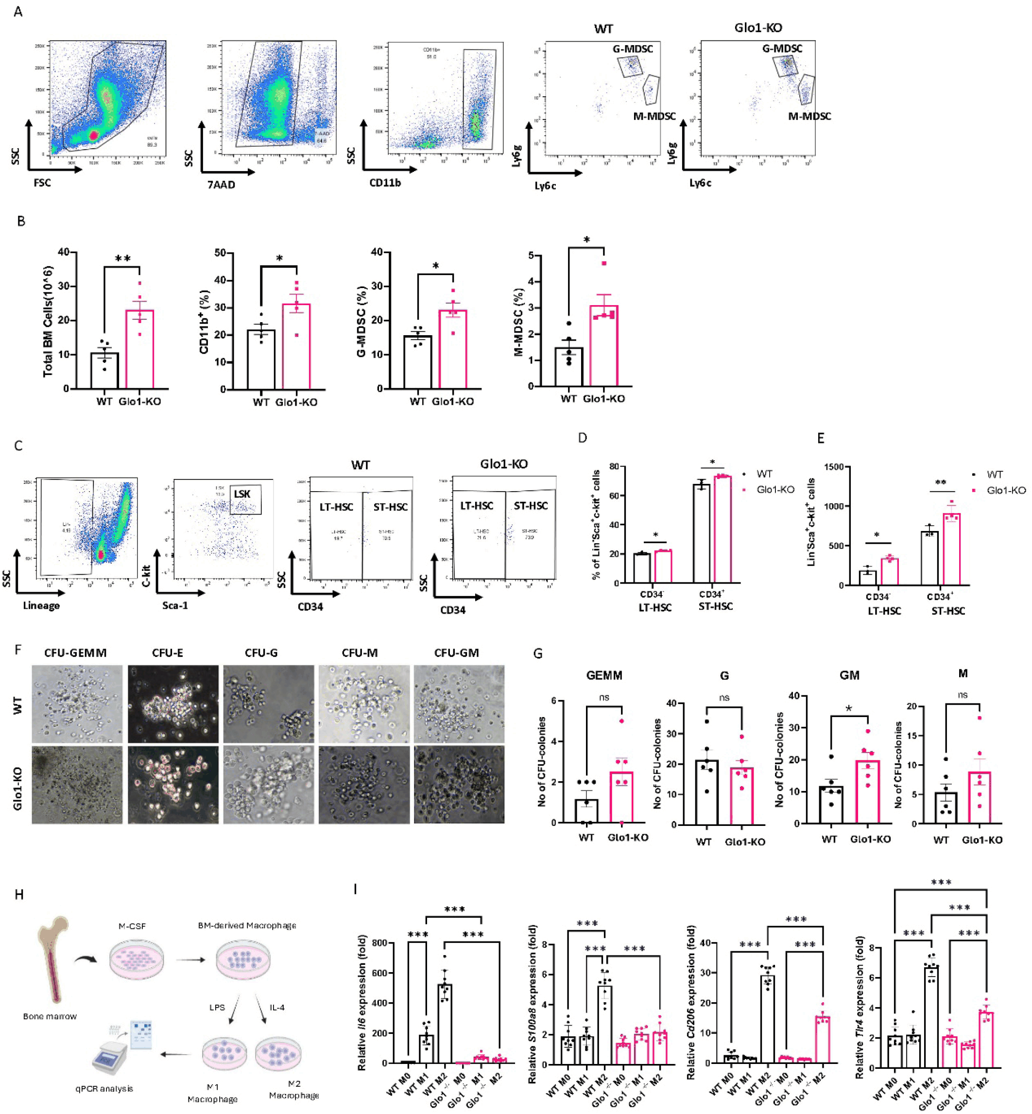
Given the observed alterations in the BM myeloid compartment, we next assessed whether GLO1 deficiency was associated with changes in the polarization responses of BM-derived macrophages. Macrophages differentiated from WT and Glo1-KO BM cells were polarized toward M1 or M2 phenotypes, and the expression of macrophage polarization-associated genes was subsequently examined (Fig. 1H). Despite the expansion of BM myeloid and hematopoietic stem/progenitor compartments, Glo1-KO-derived macrophages showed attenuated expression of Il6, S100a8, and Cd206 in response to the respective polarization conditions, together with altered Tlr4 expression (Fig. 1I). The observed changes are consistent with a myeloid-biased defined hematopoietic stem/progenitor populations and altered macrophage responsiveness to polarization stimuli.
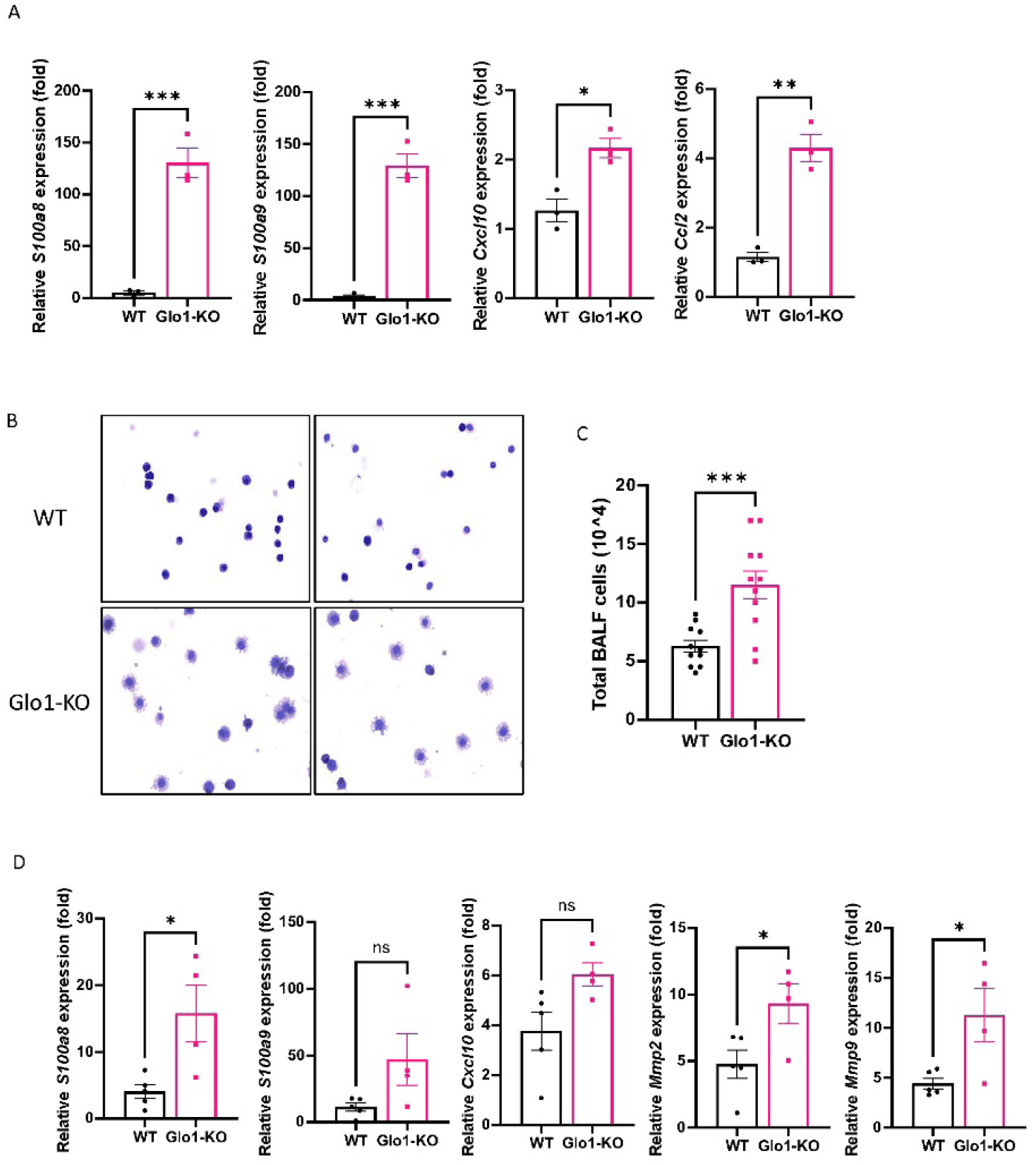
Given the myeloid expansion and altered macrophage responsiveness observed in the BM of Glo1-KO mice, we next assessed whether these alterations were reflected in peripheral inflammatory response. In peripheral blood mononuclear cells (PBMCs), Glo1-KO mice exhibited markedly increased expression of S100a8, S100a9, Cxcl10, and Ccl2 compared with WT mice (Fig. 2A). We further examined cellular accumulation in the airways by analyzing BALF. Glo1-KO mice exhibited a significant increase in total BALF cell numbers compared with WT mice (Fig. 2B and C), suggesting enhanced cellular recruitment or retention in the lung. Morphological assessment of BALF cells revealed an enlarged cellular morphology with increased cytoplasmic area in Glo1-KO mice compared with WT mice. Accumulated cells predominantly showed a large mononuclear, macrophage/monocyte-like morphology with abundant cytoplasm, although cytospin morphology alone could not distinguish resident alveolar macrophages from recruited monocytes. Together with the increased BALF cellularity, these morphological changes reflected an altered cellular state in the pulmonary environment. Lung tissues from Glo1-KO mice also showed significantly increased expression of the inflammatory and tissue-remodeling-associated genes S100a8, Mmp2, and Mmp9, whereas changes in S100a9 and Cxcl10 showed an increasing trend without reaching statistical significance. Overall, GLO1 deficiency was associated with inflammatory alteration extending from the circulation of the lung, accompanied by increased pulmonary cellularity and enhanced expression of tissue-remodeling-associated genes. These coordinated changes support the presence of a dysregulated peripheral immune environment in Glo1-KO mice.
DISCUSSION
Immune homeostasis is shaped by continuous interactions between hematopoietic and peripheral tissue environments, and disruption of this balance can alter both myeloid-cell composition and inflammatory responses. Experimental studies have shown that disruption of hematopoietic regulation can reshape lineage output and promote myeloid-biased hematopoiesis. Fulzele et al. demonstrated that osteocyte-specific loss of Gsα altered the BM environment and caused marked expansion of myeloid cells in the BM, spleen, and peripheral blood, indicating that changes in the surrounding niche can directly influence myelopoiesis (Fulzele et al., 2013). More recently, inflammatory remodeling of BM stromal compartments was shown to modify local monocyte dynamics through altered stromal chemokine production (Swann et al., 2026). These findings are consistent with broader evidence that hematopoietic stem cell and progenitor cell behavior is strongly influenced by signals from the surrounding niche (Pinho & Frenette, 2019). MDSC populations are likewise known to expand under a range of pathological conditions, including cancer and obesity, where they can influence immune regulation and disease progression (Wang et al., 2016; Turbitt et al., 2019).
In Glo1-KO mice, the concomitant increase in BM cellularity, CD11b+ cells, G-MDSCs, and M-MDSCs points to a broad reorganization of the myeloid compartment rather than expansion of a single population. This myeloid-biased phenotype was accompanied by increased frequencies and absolute numbers of phenotypically defined LT-HSC and ST-HSC populations. CFU analysis further revealed a selective increase in CFU-GM formation, whereas other colony types were not significantly changed, suggesting that GLO1 deficiency is associated with selective changes in granulocyte-macrophage colony-forming activity rather than a generalized increase in clonogenic output. BM-derived macrophages from Glo1-KO mice also exhibited altered transcriptional responses to polarization stimuli, suggesting that the hematopoietic changes associated with GLO1 deficiency extend beyond cell abundance to myeloid functional responsiveness. This combination of myeloid-biased hematopoiesis expanded phenotypic stem/progenitor populations, increased CFU-GM formation, and altered macrophage responsiveness is consistent with broader dysregulation of myeloid homeostasis in the absence of GLO1.
The peripheral phenotype observed in Glo1-KO mice was characterized by inflammatory changes in circulating immune cells together with cellular and molecular alterations in the lung. Elevated S100a8 and S100a9 expressions in PBMCs are of particular interest, as S100a8/A9 has been implicated in pulmonary inflammatory responses and neutrophil recruitment in experimental lung injury (Kuipers et al., 2013). Increased Ccl2 and Cxcl10 expressions may also reflect enhanced chemotactic signaling, as both chemokines have been shown to regulate leukocyte recruitment and inflammatory cell accumulation in the lung (Maus et al., 2002; Jiang et al., 2012). These changes were accompanied by increased BALF cellularity and elevated pulmonary expression of S100a8, Mmp2, and Mmp9. Notably, MMP2 and MMP9 are frequently increased during experimental lung injury and fibrosis and are closely associated with extracellular matrix remodeling, although their direct contribution to fibrogenesis may vary depending on the context of the disease (Bormann et al., 2022). Taken together, the concurrent changes in circulating inflammatory mediators, airway cellularity, and remodeling-associated genes indicate that the peripheral phenotype of Glo1-KO mice involves both inflammatory and tissue-remodeling features.
The present study extends the biological relevance of GLO1 beyond its established role in metabolic and oxidative stress by showing that GLO1 deficiency is accompanied by coordinated changes in hematopoietic and peripheral immune compartments. The use of a Glo1-KO model allowed these alterations to be examined at the host level: however, this approach also limits our ability to distinguish direct effects of GLO1 loss in immune cells from secondary effects arising from changes in the surrounding tissue environment. In addition, the expanded G-MDSC and M-MDSC populations were defined phenotypically, and their suppressive activity was not directly assessed. Similarly, macrophage responses were evaluated primarily at the transcriptional level, and the functional consequences of these changes remain to be determined. Further studies using cell-specific Glo1 deletion and functional analyses of myeloid populations will be required to define the mechanisms linking GLO1 deficiency to the inflammatory and tissue-remodeling phenotypes observed in peripheral tissues.


