ARTICLE

Modulation of Neural Circuit Actvity by Ethanol in Basolateral Amygdala

Leeyup Chung†
Author Information & Copyright
Corresponding author: Leeyup Chung, 595 LaSalle St, GSRB1 4023, Box 103856, Durham NC, 27710 USA. Phone: +1-919-613-5104, E-mail: leeyup@duke.edu

Copyright © 2012 © Korean Society of Developmental Biology. All Rights Reserved. This is an Open Access article distributed under the terms of the Creative Commons Attribution Non-Commercial License (http://creativecommons.org/licenses/by-nc/3.0/) which permits unrestricted non-commercial use, distribution, and reproduction in any medium, provided the original work is properly cited.

Received: Oct 3, 2012 ; Revised: Nov 23, 2012 ; Accepted: Nov 27, 2012

ABSTRACT

Ethanol actions in the amygdala formation may underlie in part the reinforcing effects of ethanol consumption. Previously a physiological phenomenon in the basolateral amygdala (BLA) that is dependent on neuronal network activity, compound postsynaptic potentials (cPSPs) were characterized. Effects of acute ethanol application on the frequency of cPSPs were subsequently investigated. Whole cell patch clamp recordings were performed from identified projection neurons in a rat brain slice preparation containing the amygdala formation. Acute ethanol exposure had complex effects on cPSP frequency, with both increases and decreases dependent on concentration, duration of exposure and age of the animal. Ethanol produces complex biphasic effects on synaptically-driven network activity in the BLA. These findings may relate to subjective effects of ethanol on arousal and anxiolysis in humans.


INTRODUCTION

A significant component of the reinforcing properties of ethanol may be its pronounced anxiolytic effect, and individuals with clinically diagnosed anxiety disorders are at greatly increased risk for ethanol abuse and dependence. The amygdala formation has a critical role in the action of anxiolytic drugs such as ethanol, and is likely to be an important brain area for investigating the mechanisms of regulation of anxiety by ethanol.

In the basolateral amygdala (BLA), ethanol inhibits projection neuron activity in vivo (Perra et al., 2008). In the BLA in vitro preparation, ethanol increases spontaneous and evoked GABA release (Zhu & Lovinger 2006; Silberman et al., 2008), inhibits glutamatergic transmission (Gean 1992; Calton et al., 1998; Lack et al., 2008) and inhibits voltage-dependent calcium channels (Calton et al., 1999). However, studies of ethanol effects on recurring neuronal circuit activity in brain areas such as the amygdala are rare. In the BLA, compound postsynaptic potentials (cPSPs) represent spontaneous circuit activity that requires both GABA and glutamate neurotransmission (Smith & Dudek, 1996; Rainnie, 1999). It was recently demonstrated that the frequency of cPSPs recorded in vitro in BLA is modulated in a consistent fashion by neuropeptides known to regulate anxiety states (Chung & Moore, 2009). Because ethanol also modulates glutamate and GABA neurotransmission, whether cPSPs might be modulated by ethanol was investigated using a whole cell patch recording technique in a rat brain slice preparation containing the amygdala formation. Using cPSPs as a model, it was attempted to characterize ethanol effects on neuronal circuit activity in BLA. Since ethanol is known to have age-dependent effects on anxiety-related behaviors in rodents (Silveri & Spear, 1998; Varlinskaya and Spear, 2002a, 2002b), effects of ethanol on cPSPs recorded from slices from immature and mature rats was also investigated.

MATERIALS AND METHODS

1. Brain slices

All procedures were approved by the Duke University and Durham VAMC Animal Care and Use Committees. Male Sprague-Dawley rats (16 to 23 or 75 to 79 days old) were decapitated under isoflurane anesthesia. Brains were quickly removed and placed into a chilled solution containing (in mM) 2.5 KCl, 1.25 NaH2PO4, 10.0 MgSO4, 0.5 CaCl2, 26.0 NaHCO3, 11.0 glucose, and 234.0 sucrose bubbled with 95% O2/5% CO2. Coronal slices (300 μm) containing the amygdala were made using a vibrating tissue slicer. The slices were incubated for 30 minutes at 35°C in artificial cerebrospinal fluid (ACSF) containing (in mM) 126.0 NaCl, 2.5 KCl, 1.25 NaH2PO4, 1.0 MgSO4, 2.0 CaCl2, 26.0 NaHCO3, and 10.0 glucose bubbled with 95% O2/5% CO2. After another 1 hour at room temperature, a single slice was transferred to the recording chamber.

2. Whole cell recording

Individual cells were visualized using a microscope (Axioskop2, Zeiss). Whole cell recordings were obtained in current clamp mode using a glass micropipette (1.5 mm OD, WPI) containing (in mM) 130 K-gluconate, 10 KCl, 10 HEPES, 2 MgCl2, 2 Mg-ATP, 0.2 Tris-GTP, 0.5 EGTA, and 5 phosphocreatinine. Temperature was maintained at 30°C. Signals were amplified using an AxoPatch 200B (Axon Instrument) and recorded onto the hard drive of an IBM-compatible computer. The calculated junction potentials (pClamp 10) were approximately 14 mV, but were not compensated in the reported membrane potential. All drugs were bath applied in ACSF with about 1 minute delay to reach the recording chamber. The cPSP events were detected with Minianalysis (Synaptosoft). To calculate the cPSP frequency ratio, the frequency at each minute was normalized to the baseline frequency (the mean cPSP frequency for the initial 5 or 10 minutes prior to experimental treatments). One-way repeated-measures ANOVA was used to test the main effect and the significance at each minutes compared with baseline value. Statistical significance was set at p<0.05 (Prism 4, GraphPad). All drugs were from Sigma (Sigma-Aldrich, St. Louis, MO).

RESULTS

1. Ethanol modulates cPSP frequency in BLA slices from young animals

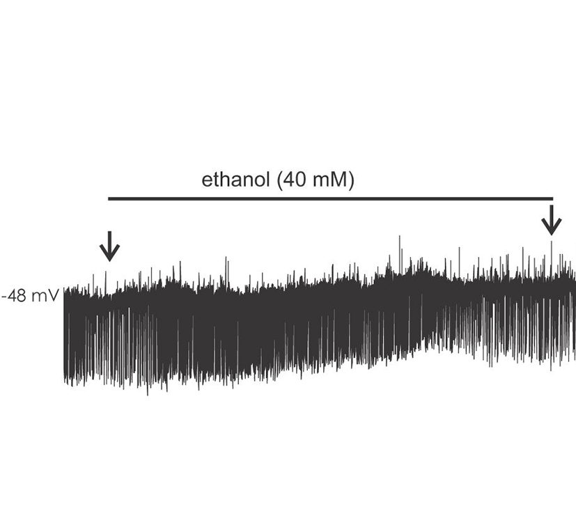
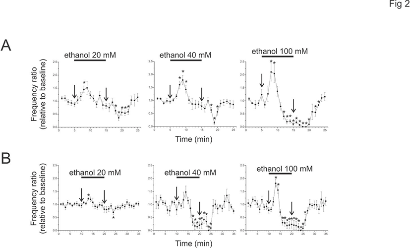
Ethanol modulated the circuit activity in BLA in a dose and time dependent manner. cPSPS in BLA was examined in slices from 16-23 days old rats (P16-23) (base cPSP frequency mean, 22.1±4.9; range, 8.8-49.6/minute, n=9). Acute application of 20 mM ethanol significantly increased the initial frequency ratio of cPSPs within 3 minutes (at 3 minutes after the application, 1.45±0.12). The frequency ratio of cPSPs subsequently decreased during washout (at 4 minutes after washout, 0.36±0.11). At 40 mM (base frequency mean, 29.3±4.2; range, 12.4-51.6/minute, n=10), the increase in frequency ratio cPSP was more pronounced (1.61±0.14), as was the decrease after washout (0.15±0.06). At 100 mM ethanol (base frequency mean 23.3±3.6; range, 10.2-33.6/minute, n=6), an initial frequency ratio increase was briefly present (2.12±0.35), but was followed by a strong attenuation (0.22±0.08) with a gradual return to baseline upon washout. Fig. 1.
dr-16-4-265-g001
Fig. 1. Ethanol modulates cPSPs in basolateral amygdala slices. A. An example of ethanol effect (40 mM) from 21 days old rat. The cPSP frequency increases after 2 to 3 minutes of ethanol application (total duration, 10 minutes). After washout for 4 minutes, cPSP frequency is decreased transiently. The begining and end of the application are indicated by two arrows. The membrane potential was depolarized (–48 mV) because of 5 mM extracellular K+.
Download Original Figure
2. Ethanol modulates cPSP frequency in BLA slices from adult animals

To test whether a similar ethanol effect is present in adult animals, ethanol of 20, 40, or 100 mM was applied to brain slices from P75-79 (Fig. 2). The cPSPs were present in adult animals (10 minute baseline, 20 mM, mean 36.7±4.1, range 14.2-55.3/minute, n=9; 40 mM, mean 33.2±6.4, range 19.6-54.2/minute, n=5; 100 mM, mean 23.8±2.2, range 16.6-28.4/minute, n=5). The peak enhancement of cPSP frequency ratio was larger than baseline at 20 and 100 mM (20 mM, 1.2±0.0; 100 mM, 1.82±0.15), but was not at 40 mM (1.48±0.23). However, during the ethanol application the frequency ratio was decreased at 40 and 100 mM (40 mM, 0.15±0.08 at 9 minutes after ethanol; 100 mM, 0.21±0.15), but not at 20 mM (0.82±0.12 at 10 minutes after ethanol). At 4 minutes after the start of washout, cPSPs were decreased at 20 mM and almost absent at 40 and 100 mM (20 mM, 0.70±0.01; 40 mM, 0.01±0.01; 100 mM, 0.01±0.01).
dr-16-4-265-g002
Fig. 2. Ethanol effects on cPSPs in slices from young and adult animals. A. The cPSP frequency ratio changes from young animals (P16-23). At 20 mM, a stimulatory effect is evident (n=9). At 40 mM, an initial increase is followed by a transient decrease during washout (n=10). At 100 mM, the transient increase is followed by strong inhibition during ethanol application (n=6). Ethanol was applied only once to each slices for 10 minutes as indicated by two arrows. The cPSP frequency is normalized to 5 minutes of baseline recording just before ethanol application. Asterisks ("*"") indicate the significant differences at the time relative to the baseline. The p-values of 20 mM ethanol are 0.008 (8 min), 0.013 (18 min), 0.000 (19 min), 0.005 (20 min), 0.001 (21 min) and 0.003 (22 min). The p-values of 40 mM ethanol are 0.002 (8 min), 0.002 (9 min), 0.012 (10 min), 0.006 (18 min), 0.000 (19 min) and 0.010 (20 min). The p-values of 100 mM are 0.023 (8 min), 0.027 (9 min), 0.003 (12 min), 0.000 (13 min), 0.000 (14 min), 0.000 (15 min), 0.000 (16 min), 0.000 (17 min), 0.000 (18 min), 0.000 (19 min), 0.000 (20 min), 0.230 (21 min), 0.030 (22 min) and 0.005(23 min). B. The cPSP frequency ratio changes from adult animals (P75-78). Ethanol at 20 mM increases cPSPs only slightly (n=9). Ethanol at 40 mM shows weak initial stimulation followed by strong inhibition (n=5). Ethanol at 100 mM shows initial stimulation followed by strong inhibition (n=5). The cPSP frequency is normalized to 10 minutes of baseline just before ethanol application. The p-values at 20 mM ethanol are 0.002 (13 min) and 0.017 (24 min). The p-values at 40 mM ethanol are 0.007 (17 min), 0.004 (18 min), 0.000 (19 min), 0.004 (20 min), 0.010 (21 min), 0.028 (22 min), 0.030 (23 min), 0.000 (24 min) and 0.000 (25 min). The p-values at 100 mM ethanol are 0.005 (13 min), 0.013 (14 min), 0.002 (16 min), 0.004 (17 min), 0.002 (18 min), 0.006 (19 min), 0.006 (20 min), 0.012 (21 min), 0.008 (22 min), 0.003 (23 min), 0.000 (24 min), 0.000 (25 min) and 0.049 (26 min).
Download Original Figure

DISCUSSION

A recent work suggests that endogenous neuropeptides may modulate cPSP frequency in BLA, with anxiogenic peptides enhancing cPSP frequency and anxiolytic peptides reducing it (Chung & Moore, 2009). The current data indicate that ethanol has dose and duration effects on cPSP frequency that include both stimulation and inhibition. Biphasic effects of ethanol have been previously reported in humans and animals (Addicott et al., 2007; Pohorecky, 1977). In rats, ethanol injection both increased and decreased the release of noradrenaline and acetylcholine in temporal and dose-dependent manner (Rossetti et al., 1992; Stancampiano et al., 2004). The stimulating effect of ethanol may reflect the predisposition to alcohol addiction (Beckstead and Phillips 2009; Meyer et al., 2009). In this regard, it is important to note that ethanol stimulates dopaminergic neurons in ventral tegmental area (Okamoto et al., 2006; McDaid et al., 2008). Our data indicate that similar phenomenon may be present in BLA. In brain slices from young animals, low concentrations of ethanol (e.g. 20 mM) primarily increase BLA circuit activity, perhaps leading to positive behavioral effects such as increased arousal. Higher concentrations of ethanol (e.g.>40 mM) may inhibit BLA output which may be associated with anxiolysis and sedation.

Using a brain slice preparation, a related study reported that ethanol potentiates circuit activity in the form of giant depolarizing potentials (GDPs) which depend on GABAergic depolarization in hippocampal CA3 region (Galindo et al., 2005). This study used very young animal (3 to 7 days old) and the effect was only facilitatory. The ethanol effect was absent in animals more than 2 weeks old, specifically 18 to 23 days old. However this study is different from that study in both age and effect pattern. The age was either 2-3 or 10 week old animals, and although ethanol effects on cPSPs were apparent at both time points, the effects were distinct. While the effect of ethanol in slices from young animals was biphasic, effects in older animals were primarily inhibitory. Still, it is speculated that the cPSPs in amygdala and GDPs in immature hippocampus may share some basic mechanisms, such as GABAergic depolarization.

The finding that ethanol effects on cPSPs in slices from adult rats were mainly inhibitory is consistent with a general enhancement of GABAergic tone in mature rat brains, while neuronal circuit activity in immature amygdala may respond to ethanol with dose-and time- dependent facilitation, consistent with enhanced ethanol-induced arousal in young individuals (Silveri & Spear, 1998; Varlinskaya & Spear, 2002a, 2002b). These findings in amygdala roughly parallel electrophysiological data from hippocampal slices taken from immature and adult rats (Li et al., 2003). In general, ethanol enhanced spontaneous inhibitory neurotransmission to a greater extent in slices from adult animals compared to juvenile or adolescent animals. The sensitivity of GABAA receptor-mediated inhibitory processes to ethanol might increase with development from the juvenile period to adulthood. Such a process could explain the greater arousal and less sedation produced by ethanol in younger individuals (Silveri & Spear, 1998; Varlinskaya & Spear, 2002a, 2002b; Hefner and Holmes, 2007).

REFERENCES

1.

Addicott MA, Marsh-Richard DM, Mathias CW, Dougherty DM. The biphasic effects of alcohol: comparisons of subjective and objective measures of stimulation, sedation, and physical activity. Alcohol Clin Exp Res. 2007; 31 p. 1883-1890.

2.

Beckstead MJ, Phillips TJ. Mice selectively bred for high- or low-alcohol-induced locomotion exhibit differences in dopamine neuron function. J Pharmacol Exp Ther. 2009; 329 p. 342-349.

3.

Calton JL, Wilson WA, Moore SD. Magnesium-dependent inhibition of NMDA-receptor mediated excitatory synaptic transmission by ethanol in basolateral amygdala. J Pharmacol Exp Ther. 1998; 287 p. 1015-1019.

4.

Calton JL, Wilson WA, Moore SD. Reduction of voltage-dependent currents by ethanol contributes to inhibition of NMDA receptor-mediated excitatory synaptic transmission. Brain Res. 1999; 816 p. 142-148.

5.

Chung L, Moore SD. Neuropeptides modulate compound postsynaptic postentials in basolateral amygdala. Neuroscience. 2009; p. 1389-97.

6.

Froehlich JC, Wand GS. The neurobiology of ethanol-opioid interactions in ethanol reinforcement. Alc Clin Exp Res. 1996; 20 p. 181A-186A.

7.

Froehlich J, O’Malley S, Hyytiä P, Davidson D, Farren C. Preclinical and clinical studies on naltrexone: What have they taught each other?. Alc Clin Exp Res. 2003; 273 p. 533-539.

8.

Galindo R, Zamudio PA, Valenzuela CF. Alcohol is a potent stimulant of immature neuronal networks: implications for fetal alcohol spectrum disorder. J Neurochem. 2005; 94 p. 1500-1511.

9.

Gean PW. Ethanol inhibits epileptiform activity and NMDA receptor-mediated synaptic transmission in rat amygdaloid slices. Brain Res Bull. 1992; 28 p. 417-421.

10.

Hefner K, Holmes A. An investigation of the behavioral actions of ethanol across adolescence in mice. Psychopharmacology (Berl). 2007; 191 p. 311-322.

11.

Heyser CJ, Roberts AJ, Schulteis G, Koob GF. Central administration of an opiate antagonist decreases oral ethanol self-administration in rats. Alc Clin Exp Res. 1999; 23 p. 1468-1476.

12.

Lack AK, Ariwodola OJ, Chappell AM, Weiner JL, McCool BA. Ethanol inhibition of kainite receptor-mediated excitatory neurotransmission in the rat basolateral nucleus of the amygdala. Neuropharmacology. 2008; 55 p. 661-668.

13.

Li Q, Wilson WA, Swartzwelder HS. Developmental differences in the sensitivity of hippocampal GABAA receptor-mediated IPSCS to ethanol. Alc Clin Exp Res. 2003; 27 p. 2017-2022.

14.

Li Q, Okada Y, Marczak E, Wilson WA, Lazarus LH, Swartzwelder HS. The novel µ-opioid receptor antagonist, [N-Allyl-Dmt1]endomorphin-2, attenuates the enhancement of GABAergic neurotransmission by ethanol. Alcohol & Alcoholism. 2009; 44 p. 13-19.

15.

McDaid J, McElvain MA, Brodie MS. Ethanol effects on dopaminergic ventral tegmental area neurons during block of Ih: involvement of barium-sensitive potassium currents. J Neurophysiol. 2008; 100 p. 1202-1210.

16.

Meyer PJ, Meshul CK, Phillips TJ. Ethanol- and cocaine-induced locomotion are genetically related to increases in accumbal dopamine. Genes Brain Behav. 2009; 8 p. 346-355.

17.

Okamoto T, Harnett MT, Morikawa H. Hyper-polarization-activated cation current (Ih) is an ethanol target in midbrain dopamine neurons of mice. J Neurophysiol. 2006; 95 p. 619-626.

18.

Perra S, Pillolla G, Luchicchi A, Pistis M. Alcohol inhibits spontaneous activity of basolateral amygdala projection neurons in the rat: involvement of the endocannabinoid system. Alcohol Clin Exp Res. 2008; 32 p. 443-449.

19.

Pohorecky LA. Biphasic action of ethanol. Biobehavioral Reviews. 1977; 1 p. 231-240.

20.

Rossetti ZL, Longu G, Mercuro G, Hmaidan Y, Gessa GL. Biphasic effect of ethanol on noradrenaline release in the frontal cortex of awake rats. Alcohol Alcohol. 1992; 27 p. 477-480.

21.

Silberman Y, Shi L, Brunso-Bechtold JK, Weiner JL. Distinct mechanisms of ethanol potentiation of local and paracapsular GABAergic synapses in the rat basolateral amygdala. J Pharmacol Exp Ther. 2008; 324 p. 251-260.

22.

Silveri MM, Spear LP. Decreased sensitivity to the hypnotic effects of ethanol early in ontogeny. Alcohol Clin Exp Res. 1998; 22 p. 670-676.

23.

Stancampiano R, Carta M, Cocco S, Curreli R, Rossetti ZL, Fadda F. Biphasic effects of ethanol on acetylcholine release in the rat prefrontal cortex. Brain Res. 2004; 997 p. 128-132.

24.

Varlinskaya EI, Spear LP. Acute effects of ethanol on social behavior of adolescent and adult rats: role of familiarity of the test situation. Alcohol Clin Exp Res. 2002a; 26 p. 1502-1511.

25.

Varlinskaya EI, Spear LP. Changes in sensitivity to ethanol-induced social facilitation and social inhibition from early to late adolescence. Ann N Y Acad Sci. 2002b; 1021 p. 459-61.

26.

Volpicelli JR, Alterman AI, Hayashida M, O'Brien CP. Naltrexone in the treatment of alcohol dependence. Arch Gen Psych. 1992; 49 p. 876-880.

27.

Vukojević V, Ming Y, D'Addario C, Rigler R, Johansson B, Terenius L. Ethanol/naltrexone interactions at the mu-opioid receptor. CLSM/FCS study in live cells. PloS ONE. 2008; 3 p. e4008.

28.

Zhu PJ, Lovinger DM. Ethanol potentiates GABAergic synaptic transmission in a postsynaptic neuron/synaptic bouton preparation from basolateral amygdala. J Neurophysiol. 2006; 96 p. 433-441.