Research articles

In Vitro Sex Steroid Metabolism in Red Spotted Grouper, Epinephelus akaara during Oocyte Maturation

In Joon Hwang1https://orcid.org/0000-0001-5740-3925, Hea Ja Baek2,https://orcid.org/0000-0002-4578-5919
Author Information & Copyright
1Inland Fisheries Research Institute, National Institute of Fisheries Science, Gapyeong 12453, Korea
2Department of Marine Biology, Pukyong National University, Busan 48513, Korea
Corresponding author Hea Ja Baek, Department of Marine Biology, Pukyong National University, Busan 48513, Korea. Tel: +82-51-629-5924, E-mail: hjbaek@pknu.ac.kr

© Copyright 2021 The Korean Society of Developmental Biology. This is an Open-Access article distributed under the terms of the Creative Commons Attribution Non-Commercial License (http://creative-commons.org/licenses/by-nc/4.0/) which permits unrestricted non-commercial use, distribution, and reproduction in any medium, provided the original work is properly cited.

Received: Mar 01, 2021 ; Revised: Apr 09, 2021 ; Accepted: May 11, 2021

Published Online: Jun 30, 2021

Abstract

We studied steroid metabolites produced from red-spotted grouper ovarian follicles during maturation. Oocytes with 350–500 μm diameter were in vitro incubated in the presence of [3H] 17α-hydroxyprogesterone as a precursor. Steroid metabolites were extracted from incubated media and oocytes. The extracts were separated and identified using thin layer chromatography, high performance liquid chromatography and gas chromatography-mass spectrometry. The identified metabolites were androstenedione (A4), testosterone (T) and estrone (E1). The metabolites of A4 was dominant in all size of oocytes and it was the highest in 480 μm diameter oocytes. The metabolites of two progestins, 17α,20β-dihydroxy-4-pregnen-3-one and 17α,20α-dihydroxy-4-pregnen-3-one were detected in the oocytes less than 480 μm diameter although they were not identified definitely. In the oocytes of 480 μm diameter, metabolite of progestin was the highest, and germinal vesicle (GV) was still in the middle of cytoplasm. In the oocytes of 500 μm diameter, GV was began to migrate and the major metabolites were A4 and E1. The metabolite of E1 was detected in all size of oocytes and it was higher than that of E2. These results suggest that oocytes of 480 μm diameter are the transitional stage involving steroidogenic shift to final oocyte maturation and potential function of E1 during maturation process.

Keywords: Red spotted grouper; Steroid metabolite; Oocyte; Maturation

INTRODUCTION

Oocyte maturation process in teleosts is under the control or feedback of sex steroids, synthesized from the follicle cell, and they are regulated by the hypothalamus-pituitary –gonad axis (Nagahama & Yamashita, 2008; Planas & Swanson, 2008). In female fish, it is well known that estradiol-17β (E2) is the major sex steroid, and transported to liver during vitellogenesis, the period of vitellogenin production in liver. Subsequently, progestins function as such as maturation inducing steroid (MIS), 17α,20β-dihydroxy-4-pregnen-3-one (17α20βP) and/or 17α,20β,21-trihydroxy-4-pregnen-3-one (17α20β21P) induce final oocyte maturation, i.e., germinal vesicle breakdown (GVBD) and ovulation (Patiño & Sullivan, 2002; Baek, 2008; Nagahama & Yamashita, 2008). Although the actions and role of these sex steroids had been studied in many fish species, the profiling of various sex steroids in fish reproduction is still insufficient because of not only diverse fish species but their diverse reproductive characteristics (Frisch A, 2004; Tokarz et al., 2015).

The red-spotted grouper, Epinephelus akaara is a marine multiple spawner and protogynous, as a Serranid fish distributed mainly in Korea, southern Japan, southern China and East China Sea (Craig et al., 2011; Lee et al., 2020). This species is economically important in aquaculture industry and it is interested in artificial control of its reproduction such as induction of maturation and masculinization (Li et al., 2006; Lee et al., 2014; Oh et al., 2018; Lee et al., 2020). However, there is little information about sex steroid synthesis related to oocyte maturation of this species. In the present study, we investigated the in vitro profiling of sex steroid metabolites from ovarian follicles of red spotted grouper.

MATERIALS AND METHODS

1. Chemicals

Authentic steroids were purchased from Sigma-Aldrich (St. Louis, MO, USA) or Steraloids (Wilton, NH, USA). Mother solutions (mg/mL) were prepared by dissolving the authentic steroids in pure ethanol. These were further diluted in incubation media. The ethanol concentration in the incubation medium was maintained at less than 0.1%. Radioactive [3H]-17α-hydroxyprogesterone ([3H]-17αP) was obtained from Amersham Life Science (London, UK).

2. Preparation of oocytes for incubation

The experimental fish were reared and adjusted to 14L:10D and 25°C in recirculating aquaria (500 L) from Marine Science Institute, Jeju National University, Korea. Oocytes were obtained by cannulation after anesthetization of eight mature females. The cannulated oocytes were put into cold balanced salt solution (BSS; 132.96 mM NaCl, 3.09 mM KCl, 0.28 mM MgSO4․7H2O, 0.98 mM MgCl2․6H2O, 3.40 mM CaCl2․6H2O, 3.65 mM HEPES). Oocytes were separated into groups with the largest diameter using fine forceps from each mature fish. Oocytes with average diameters of 350, 400, 420, 440, 480, and 500 μm were used for the in vitro incubation. Thirty follicle-enclosed oocytes were incubated in each well of triplicates.

We incubated isolated oocytes in 1 mL of Leibovitz L15 medium (Gibco, Grand Island, NY, USA) with 55 kBq [3H]-17αP a radiolabeled precursor for separation of metabolites. The pH and osmolality of the media were adjusted to 7.7 and 360 mOsm, respectively. The plates were incubated for 24 h at 18°C.

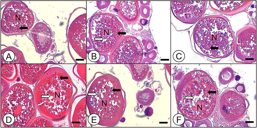
Some ovarian fragment from each fish were fixed, washed, dehydrated and paraffin-embedded. Sections of 4–5 μm were HE stained and observed through a light microscope.

3. Analysis of steroid metabolism

At the end of the incubation, steroids extraction, separation and identification of metabolites were followed by our previous studies (Hwang et al., 2012; Baek et al., 2013; Lee & Baek, 2015). Steroid metabolites produced from [3H]-17αP were compared with the values were expressed as photo-stimulated luminescence (PSL) – background (BG) per mm2 of each fraction area from autoradiography.

RESULTS

1. Oocyte developmental stage

In histological observation of each diameter of oocytes, oocytes of 350–480 μm diameter were vitellogenic stage; yolk granules were spread throughout the cytoplasm and oil droplets (Od) were accumulated near the nucleus (N) as oocyte diameter increased (Fig. 1A–E). In particular, nucleus was observed in the middle of cytoplasm in these size of oocytes. In the oocytes of 500 μm diameter, migration of N was observed and many Ods were observed near migrating N (Fig. 1F).

dr-25-2-75-g1
Fig. 1. Histological observation of ovarian fragment from mature red spotted grouper. (A) oocytes of 350 μm, (B) oocytes of 400 μm, (C) oocytes of 420 μm, (D) oocytes of 440 μm, (E) oocytes of 480 μm, (F) oocytes of 500 μm. Scale bars=100 μm, stained with H-E. N, nucleus; blank arrow, oil droplet; solid arrow, yolk granule.
Download Original Figure
2. Identification of steroid metabolites

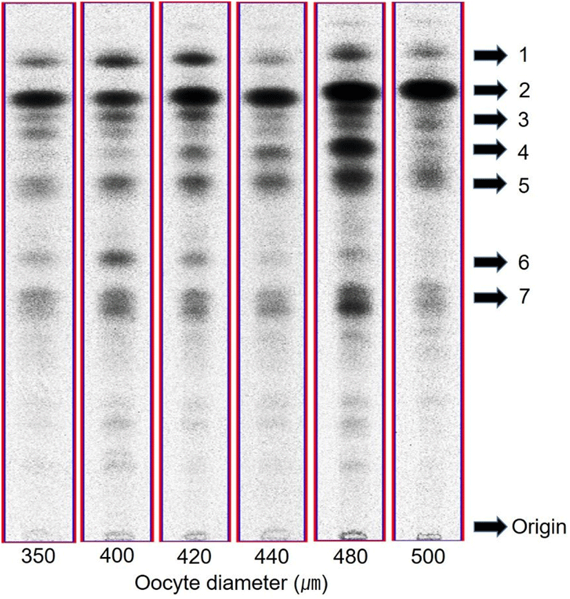
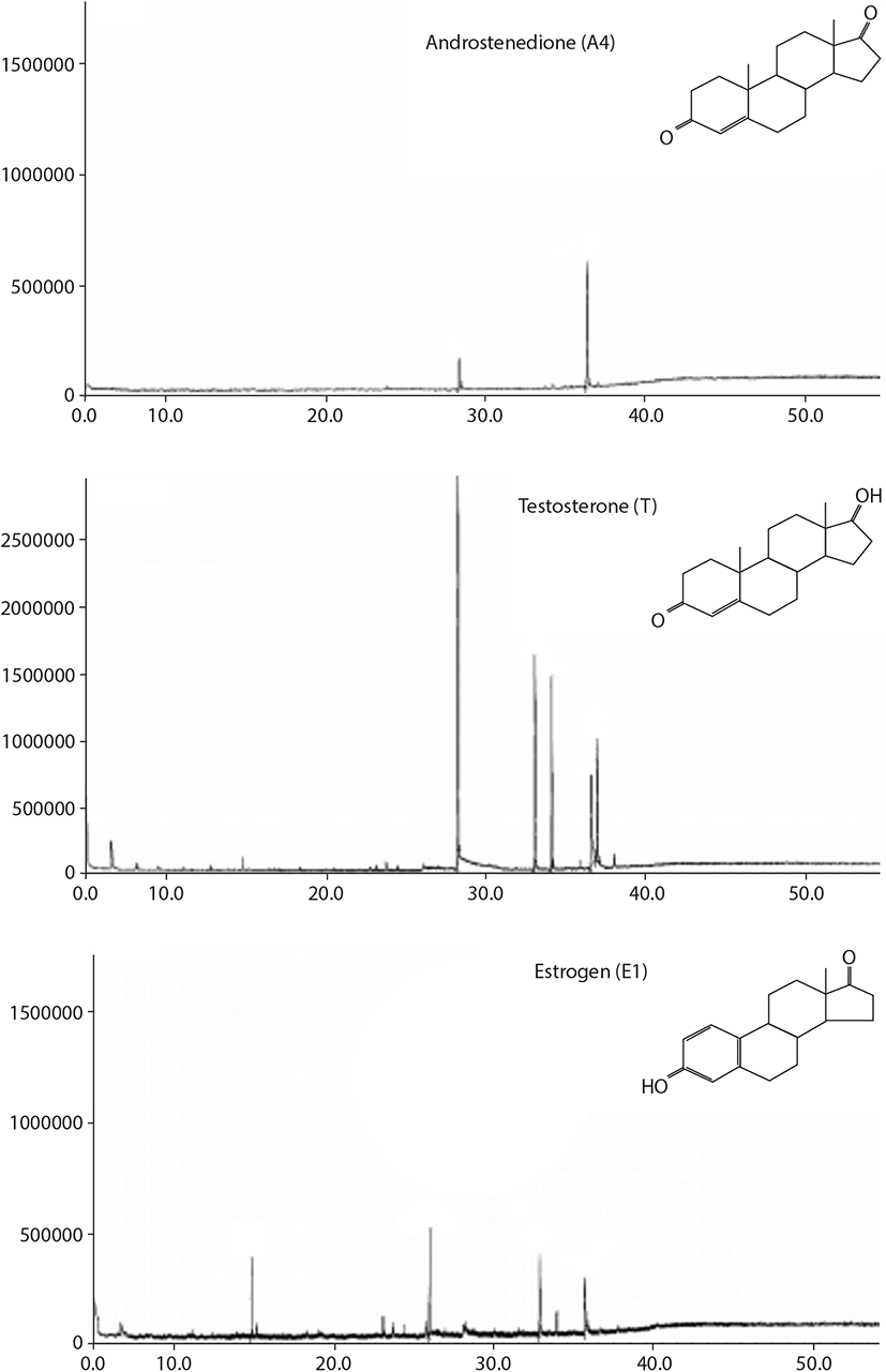
After incubation of oocytes with [3H]-17αP, 7 major fractions appeared on thin layer chromatography (TLC). Fraction 1, 2, 4, 5, and 6 were as the same retention factor (RF) of standard estrone (E1), androstenedione (A4), 17αP, T, and 17α20βP, respectively (Fig. 2). Fraction 3 and 7 were overlapped with E2+unknown metabolite and 17α,20α-dihydroxy-4-pregnen-3-one (17α20αP) +unknown metabolite, respectively. Among these 7 metabolites, fraction 1, 2, and 5 were identified as E1, A4, and T through further analysis with high performance liquid chromatography and GC/MS (Fig. 3). The other metabolites were not identified due to their low measured activities.

dr-25-2-75-g2
Fig. 2. Autoradiograms of steroid metabolites produced from [3H]17α-hydroxyprogesterone after 24 h incubation with isolated oocytes from red spotted grouper. Steroid metabolites from radiolabeled precursor were separated by thin layer chromatography developed with a benzene:acetone (4:1) and benzene:ethyl acetate (4:1) solvent system. 1, estrone (E1); 2, androstenedione (A4); 3, estradiol-17β (E2)+unknown metabolite; 4, 17αP; 5, testosterone (T); 6, 17α,20β-dihydroxy-4-pregnen-3-one (17α20βP); 7, 17α,20α-dihydroxy-4-pregnen-3-one (17α20αP)+unknown metabolite.
Download Original Figure
dr-25-2-75-g3
Fig. 3. Mass spectra of steroid metabolites identified as androstenedione, testosterone and estrone from oocytes of red spotted grouper.
Download Original Figure
3. Metabolites profiling during maturation process

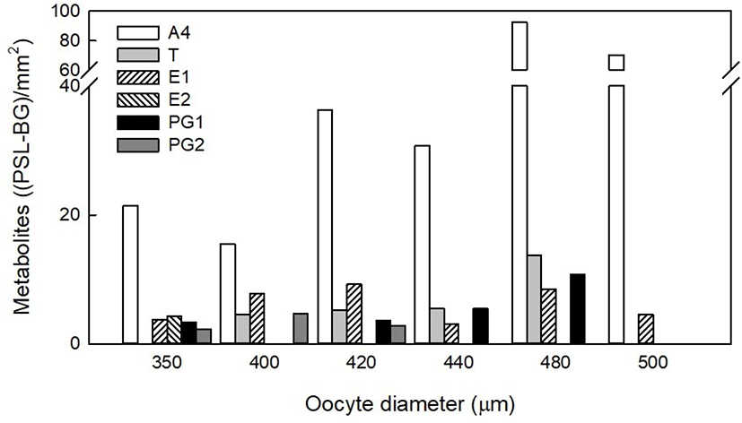
We compared each metabolite with PSL value from TLC autoradiography followed each oocyte diameter for steroid metabolites profiling during oocyte maturation (Fig. 4). The A4 metabolite was dominant [15.47–92.41 (PSL-BG)/mm2] in all size of oocytes and it was the highest in 480 μm diameter oocytes. T metabolite increased gradually [4.55–13.76 (PSL-BG)/mm2] as oocyte diameter increased although it was not detected at 350 and 500 μm diameter oocytes in the autoradiography. E2 overlapped with unknown metabolite was detected only in 350 μm diameter oocytes [4.27 (PSL-BG)/mm2]. E1 metabolite [3.08–9.24 (PSL-BG)/mm2] was detected in all size of oocytes and it was the highest in 420 μm diameter oocytes. 17α20βP metabolite was detected in 350–420 μm diameter oocytes [2.27–4.64 (PSL-BG)/mm2]. 17α20αP overlapped with unknown metabolite was detected 350, 420–480 μm diameter oocytes [3.37–10.82 (PSL-BG)/mm2] with the highest in 480 μm diameter oocytes. 17α20αP or 17α20βP metabolite was not detected in 500 μm diameter oocytes.

dr-25-2-75-g4
Fig. 4. Steroid metabolites from radioactive precursor during oocyte maturation. The values indicate digital photo-luminescence (PSL) by autoradiography and were expressed as PSL-backgroud (BG) per mm2 of each fraction area. A4, androstenedione; T, testosterone; E1, estrone; E2, estradiol-17β; PG1, 17α,20α-dihydroxy-4-pregnen-3-one; PG2, 17α,20β-dihydroxy-4-pregnen-3-one.
Download Original Figure

DISCUSSION

In general, E2, the principle estrogen is dominant in the early stage of maturation, i.e. oocyte growing and MIS such as 17α20βP or 17α20β21P dominant in the late stage of maturation, i.e. GVBD and ovulation (Tokarz et al., 2015). In histological observation of oocytes, 350–480 μm diameter oocytes were vitellogenic stage and 500 μm diameter oocytes were final maturation stage with migration of germinal vesicle (GV). We consider the 480 μm diameter oocytes would be fully vitellogenic stage; GV was still in the middle of cytoplasm and prior to GVM stage.

We identified the major steroid metabolites and investigate their profiling from vitellognic to germinal vesicle migration oocytes of red spotted grouper. The major metabolites synthesized from the oocytes were A4, E1, T, E2 and two progestins. Among these metabolites, A4, E1, and T were identified through GC/MS, but the others were not identified due to their low activities. Although we could not identify 2 progestins, progestin 1 and 2 co-migrated with 17α20αP and 17α20βP in TLC system. To date, MIS of groupers had not been identified yet although 17α20βP is well-known MIS in many fish species and was measured from plasma of Epinephelus morio (Johnson et al., 1998; Shein et al., 2004; Nagahama & Yamashita, 2008). Interestingly, progestin 1 metabolite, suspected 17α20αP increased gradually in 420–480 μm diameter oocytes. Previous studies reported that 17α20αP may act as a MIS of flatfish, Limanda limanda, Verasper variegates and gobiid fish, Chasmichthys dolichognathus (Canario & Scott, 1990; Baek, 2001, 2008). In this regard, 17α20αP metabolite would be related to maturation process with its increase until fully vitellogenic stage and then decrease at GVM stage although its role is not clear. Further study that identification of MIS and it’s maturation inducing capacity should be conducted.

Among the metabolites, A4 was predominant in all size of oocytes even in the GVM stage (500 μm oocyte diameter). In our previous study, A4 was also produced dominantly in vitellogenic oocytes from red lip mullet, Chelon haematocheilus, blacktip grouper, Epinephelus fasciatus, and blackfin flounder, Glyptocephalus stelleri (Baek et al., 2011; Hwang et al., 2012; Lee & Baek, 2015). A few studies reported that A4 is related to sex change in protogynous hermaphrodite, Monopterus albus and Sparisoma viride (Yeung & Chan, 1987; Cardwell & Liley, 1991; Yeung et al., 1993). Moreover, Montero et al. (1995) reported that androgen could act as a substrate for estrogen production in vitellogenesis of European eel, Anguilla anguilla. In the present study, we suspect that A4 may regulate oocyte maturation process rather than sex change of red spotted grouper since there was no testicular tissue in gonad and it’s the highest value with progestin production at 480 μm diameter oocytes, the transitional stage into final maturation.

We demonstrated that E1 metabolite was also detected in all size of oocytes and its detected value was higher than that of E2 metabolite. In a majority of fishes, it is well known that E2 is converted from T by aromatase (Fukada et al., 1996; Chang et al., 1997; Aggarwal et al., 2014). However, E1 may have a minor vitellogenic role in some species (Specker & Sullivan, 1994; Routledge et al., 1998) and act as a precursor of E2 synthesis (Kazeto et al., 2000; Ohta et al., 2001; Ohta et al., 2002; Mindnich et al., 2004; Zhou et al., 2005). Our results also suggest that E1 may regulate vitellogenesis with E2 and also has a certain role in oocyte maturation process despite insufficient data. Future study such as exogenous E1 treatment on oocyte maturation would provide more definite information about steroidogenic profiling. In conclusion, the present study provides in vitro steroid metabolites profiling that identification of major metabolites and its’s changes during oocytes maturation of red spotted grouper. We also suggest that 17α20βP and/or 17α20αP may act as a MIS and the potential role of E1 in maturation process.

Conflict of interests

The authors declare no potential conflict of interest.

Acknowledgements

This study was supported by a Research Grant of Pukyong National University (2019).

Authors’ contributions

Conceptualization: Baek HJ

Data curation: Hwang IJ

Formal analysis: Hwang IJ, Baek HJ

Methodology: Baek HJ

Software: Hwang IJ

Validation: Hwang IJ, Baek HJ

Investigation: Hwang IJ, Baek HJ

Writing-original draft: Hwang IJ

Writing-review & editing: Hwang IJ, Baek HJ

Ethics approval

This article does not require IRB/IACUC approval because there are no human and animal participants.

REFERENCES

1.

Aggarwal N, Goswami SV, Khandelwal P, Sehgal N. 2014; Aromatase activity in brain and ovary: Seasonal variations correlated with circannual gonadal cycle in the catfish, Heteropneustes fossilis. Indian J Exp Biol. 52:527-537.

2.

Baek HJ. 2001; Biosynthesis of 17α-hydroxy, 20α-dihydroprogesterone by ovaries of the spotted flounder (Verasper variegatus). J Fish Sci Technol. 4:70-74.

3.

Baek HJ. 2008; Production of C21 steroids in longchin goby, Chasmichthys dolichognathus during oocyte maturation. Dev Reprod. 12:107-112.

4.

Baek HJ, Hwang IJ, Lee YD, Kim HB. 2011; Effects of nonylphenol and 3,3′,4,4′,5-pentachlorobiphenyl on in vitro oocyte steroidogenesis in redlip mullet, Chelon haematocheilus. Anim Cells Syst. 15:189-196

5.

Baek HJ, Kim DG, Kim HB. 2013; In vitro steroidogenesis on oocyte development in the starry flounder, Platichthys stellatus. Dev Reprod. 17:421-426

6.

Canario AVM, Scott AP. 1990; Effects of steroids and human chorionic gonadotrophin on in vitro oocyte final maturation in two marine flatfish: The dab, Limanda limanda, and the plaice, Pleuronectes platessa. Gen Comp Endocrinol. 77:161-176

7.

Canario AVM, Scott AP. 1990; Plasma levels of ovarian steroids, including 17α-20α-dihydroxy-4-pregnen-3-one and 3β,17α,20α-trihydroxy-5β-pregnane, in female dabs (Limanda limanda)—marine flatfish—induced to mature and ovulate with human chorionic gonadotrophin. Gen Comp Endocrinol. 77:177-191

8.

Cardwell JR, Liley NR. 1991; Hormonal control of sex and color change in the stoplight parrotfish, Sparisoma viride. Gen Comp Endocrinol. 81:7-20

9.

Chang XT, Kobayashi T, Kajiura H, Nakamura M, Nagahama Y. 1997; Isolation and characterization of the cDNA encoding the tilapia (Oreochromis niloticus) cytochrome P450 aromatase (P450arom): Changes in P450arom mRNA, protein and enzyme activity in ovarian follicles during oogenesis. J Mol Endocrinol. 18:57-66

10.

Craig MT, Sadovy de Mitcheson YJ, Heemstra PC. 2011 Groupers of the World: A Field and Market Guide.National Inquiry Services Center [NISC]. Grahamstown, South Africa: .

11.

Frisch A. 2004; Sex-change and gonadal steroids in sequentially-hermaphroditic teleost fish. Rev Fish Biol Fish. 14:481-499

12.

Fukada S, Tanaka M, Matsuyama M, Kobayashi D, Nagahama Y. 1996; Isolation, characterization, and expression of cDNAs encoding the medaka (Oryzias latipes) ovarian follicle cytochrome P-450 aromatase. Mol Reprod Dev. 45:285-290

13.

Hwang IJ, Kim SK, Choi SJ, Lee CH, Lee YD, Kim HB, Baek HJ. 2012; Effects of steroid hormones on in vitro GVBD and oocyte steroidogenesis in blacktip grouper, Epinephelus fasciatus. Dev Reprod. 16:39-45.

14.

Johnson K, Thomas P, Wilson RR. 1998; Seasonal cycles of gonadal development and plasma sex steroid levels in Epinephelus morio, a protogynous grouper in the eastern Gulf of Mexico. J Fish Biol. 52:502-518

15.

Kazeto Y, Ijiri S, Matsubara H, Adachi S, Yamauchi K. 2000; Cloning of 17β-hydroxysteroid dehydrogenase-I cDNAs from Japanese eel ovary. Biochem Biophys Res Commun. 279:451-456

16.

Lee CH, Hur SW, Kim BH, Soyano K, Lee YD. 2020; Induced maturation and fertilized egg production of the red-spotted grouper, Epinephelus akaara, using adaptive physiology of photoperiod and water temperature. Aquac Res. 51:2084-2090

17.

Lee CH, Hur SW, Na OS, Baek HJ, Noh CH, Han SH, Lee YD. 2014; Induction of primary male in juvenile red spotted grouper Epinephelus akaara by immersion of 17α-methyltestosterone. Dev Reprod. 18:127-131

18.

Lee HW, Baek HJ. 2015; Steroid metabolism in the blackfin flounder Glyptocephalus stelleri during oocyte maturation. Korean J Fish Aquat Sci. 48:483-488

19.

Li GL, Liu XC, Lin HR. 2006; Effects of aromatizable and nonaromatizable androgens on the sex inversion of red-spotted grouper (Epinephelus akaara). Fish Physiol Biochem. 32:25-33

20.

Mindnich R, Deluca D, Adamski J. 2004; Identification and characterization of 17β-hydroxysteroid dehydrogenases in the zebrafish, Danio rerio. Mol Cell Endocrinol. 215:19-30

21.

Montero M, Le Belle N, King JA, Millar RP, Dufour S. 1995; Differential regulation of the two forms of gonadotropin-releasing hormone (mGnRH and cGnRH-II) by sex steroids in the European female silver eel (Anguilla anguilla). Neuroendocrinology. 61:525-535

22.

Nagahama Y, Yamashita M. 2008; Regulation of oocyte maturation in fish. Dev Growth Differ. 50:S195-S219

23.

Oh SB, Lee CH, Lee YD. 2018; Induction of puberty in red spotted grouper, Epinephelus akaara by water temperature. J Aquac Res Dev. 9:1000537

24.

Ohta K, Mine T, Yamaguchi A, Matsuyama M. 2001; Steroidogenic pathway to estradiol-17β synthesis in the ovarian follicles of the protogynous wrasse, Pseudolabrus sieboldi. Zool Sci. 18:937-945

25.

Ohta K, Yamaguchi S, Yamaguchi A, Okuzawa K, Kagawa H, Matsuyama M. 2002; Biosynthesis of estradiol-17β in the ovarian follicles of the red seabream Pagrus major during vitellogenesis. Fish Sci. 68:680-687

26.

Patiño R, Sullivan CV. 2002; Ovarian follicle growth, maturation, and ovulation in teleost fish. Fish Physiol Biochem. 26:57-70.

27.

Planas JV, Swanson P. 2008; Physiological function of gonadotropins in fish. In: Rocha MJ, Arukwe A, Kapoor BG, editors. Fish Reproduction. Science Publishers. Enfield, NH: p. 37-66.

28.

Routledge EJ, Sheahan D, Desbrow C, Brighty GC, Waldock M, Sumpter JP. 1998; Identification of estrogenic chemicals in STW effluent. 2. In vivo responses in trout and roach. Environ Sci Technol. 32:1559-1565

29.

Shein NL, Chuda H, Arakawa T, Mizuno K, Soyano K. 2004; Ovarian development and final oocyte maturation in cultured sevenband grouper Epinephelus septemfasciatus. Fish Sci. 70:360-365

30.

Specker JL, Sullivan CV. 1994; Vitellogenesis in fishes: Status and perspectives. In: Davey KG, Peter RE, Tobe SS, editors. Perspectives in Comparative Endocrinology. National Research Council of Canada. Ottawa, ON: p. 304-315.

31.

Tokarz J, Möller G, Hrabě de Angelis M, Adamski J. 2015; Steroids in teleost fishes: A functional point of view. Steroids. 103:123-144

32.

Yeung WSB, Chan STH. 1987; The plasma sex steroid profiles in the freshwater, sex-reversing teleost fish, Monopterus albus (Zuiew). Gen Comp Endocrinol. 65:233-242

33.

Yeung WSB, Chen H, Chan STH. 1993; The in vitro metabolism of radioactive androstenedione and testosterone by the gonads of the protogynous Monopterus albus at different sexual phases: A time-course and seasonal study. Gen Comp Endocrinol. 89:313-322

34.

Zhou LY, Wang DS, Senthilkumaran B, Yoshikuni M, Shibata Y, Kobayashi T, Sudhakumari CC, Nagahama Y. 2005; Cloning, expression and characterization of three types of 17β-hydroxysteroid dehydrogenases from the Nile tilapia, Oreochromis niloticus. J Mol Endocrinol. 35:103-116