


INTRODUCTION
In jawed fish, facial cartilages provide the framework for the jaw and branchial basket skeleton, which are key structures for respiration and feeding (Donoghue et al., 2006; DeLaurier, 2019). During embryogenesis, they arise from the pharyngeal arches, the embryonic structures composed of the cranial neural crest-derived ectomesenchyme; the jaw and jaw-support cartilaginous elements of the jaw develop from the first and second arches, and the gill-support cartilaginous elements of the branchial basket form from the posterior arches (DeLaurier, 2019). The cartilaginous elements in each arch are segmented dorsoventrally, articulated, and eventually ossify during larval stages (DeLaurier, 2019). Genetic mechanisms underlying the development of the jaw and jaw-support cartilaginous elements are well understood and largely conserved among each other, whereas those for the development of branchial basket elements are relatively less understood (DeLaurier, 2019). Recent studies in zebrafish are revealing the genetic and developmental bases for the cartilaginous elements of the branchial basket, which are more derived than previously thought (Jeon et al., 2025; Choe et al., 2026). Here, we review the developmental genetic processes for facial cartilage formation in the jaw and branchial basket of jawed fish, specifically highlighting recent findings on the unique characteristics of dorsal cartilage development in the zebrafish branchial basket.
FACIAL CARTILAGE ELEMENTS OF JAWED FISH
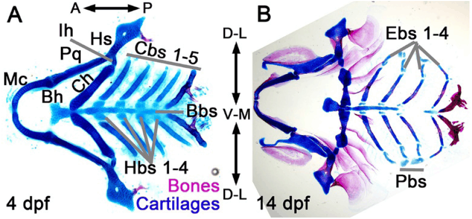
In jawed fish, the jaw, jaw support, and branchial basket are initially organized with a series of cartilaginous elements (Fig. 1). From ventral to dorsal, in the first arch, the paired Meckel’s and palatoquadrate cartilages form the jaw; in the second arch, the unpaired median basihyal, followed by the paired ceratohyal, interhyal, and hyosymplectic cartilages form the jaw support; in the posterior arches, the unpaired median basibranchial, followed by the paired hypobranchials, ceratobranchials, epibranchials, and pharyngobranchials, forms the branchial basket (Schilling & Kimmel, 1997; Pradel et al., 2014). Developmental genetic studies in zebrafish strongly suggest that, in the jaw and jaw-support structures, the Meckel’s and ceratohyal cartilages are serial ventral homologs and the palatoquadrate and hyosymplectic cartilages are serial dorsal homologs (Schilling & Kimmel, 1997; Medeiros & Crump, 2012; Barske et al., 2016). In the branchial basket, it is suggested that the median basibranchial cartilages are serial ventral homologs with the basihyal cartilage of the jaw-support structure, the ceratobranchial cartilages are serial ventral homologs with the Meckel’s and ceratohyal cartilages of the jaw and jaw-support structures, and the epibranchial cartilages are serial dorsal homologs with the palatoquadrate and hyosymplectic cartilages of the jaw and jaw-support structures (Schilling & Kimmel, 1997). However, a series of homologs for the ventral hypobranchial and dorsal pharyngobranchial cartilages of the branchial basket are not clearly distinguished between the jaw and jaw-supporting elements. Facial cartilage elements of jawed fish and their serial homology are summarized in Table 1.
FACIAL CARTILAGE DEVELOPMENT IN JAWED FISH
During neurulation, the neural crest forms on the dorsal areas adjacent to the neural tube. In the adjacent regions to the anterior neural tube in which the midbrain and hindbrain develop, a population of neural crest, the cranial neural crest, is delaminated and migrates ventrally into the pharyngeal areas as the three trigeminal, hyoid, and branchial streams to fill the first, second, and third pharyngeal arches, respectively (Knight & Schilling, 2006; Fig. 2A). During migration and after entering the pharyngeal arches, cranial neural crest cells differentiate into the ectomesenchymal cells from which cartilage, bone, and connective tissue arise (Blentic et al., 2008). Genetically, neural crest, including the cranial neural crest, is marked with transcription factors such as Sox10 and Foxd3, whereas ectomesenchyme is labeled with transcription factors such as Dlx2 and Twist1 (Blentic et al., 2008; Cox et al., 2012; Fig. 2A and B). While the first and second arches and the second and third arches are physically separated by the first and second pharyngeal pouches that are the outgrowth of the pharyngeal endoderm, the third arch is sequentially segmented into a series of posterior arches by a series of pharyngeal pouches (Choe et al., 2013; Fig. 2B). Amorphic or hypoplastic pouches result in fusion of pharyngeal arches, consequently leading to fusion among cartilaginous elements arising in each arch (Choe & Crump, 2014).
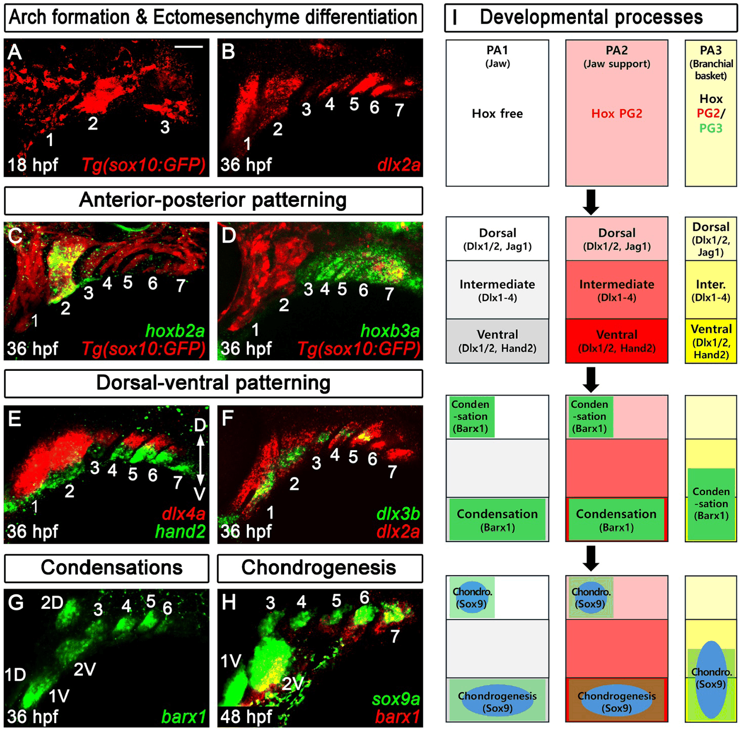
The cartilaginous elements of the jaw, jaw support, and branchial basket originate from the first, second, and posterior arches. Although they are formed from similar ectomesenchymal tissues, the serially homologous cartilaginous elements exhibit distinct shapes. This is because the pharyngeal arches are regionalized along the anterior-posterior axis by the pharyngeal Hox code. In jawed fish, the first arch is negative for Hox expression, whereas the second and third arches are positive for Hox paralogue group 2 and Hox paralogue groups 2 and 3, respectively, with the fourth and more posterior arches being for Hox paralogue groups 2 to 4 and various combinations of Hox paralogue groups, respectively (Schilling et al., 2001; Hunter & Prince, 2002; Minoux & Rijli, 2010; Oulion et al., 2011; Fig. 2C, D, and I). The pharyngeal Hox code is established through the anterior-posterior patterning of the hindbrain; as reiterated rhombomeres in the hindbrain initiate spatially restricted Hox expression via retinoic acid gradients, cranial neural crest cells adjacent to the hindbrain migrate ventrally into the arches while retaining positional memory (Schilling & Knight, 2001). In zebrafish, ectopic expression of Hox paralogue group 2 genes causes jaw-support-like cartilaginous elements instead of jaw elements in the first arch, whereas knockdown of Hox paralogue group 2 genes leads to jaw-like cartilaginous elements instead of jaw-support elements in the second arch (Hunter & Prince, 2002).
In each pharyngeal arch, the segmented ventral and dorsal cartilaginous elements for the jaw, jaw support, and branchial basket form. In jawed fish, the pharyngeal arches are regionalized into the ventral, intermediate, and dorsal areas via the nested dlx expression, the Dlx code (Medeiros & Crump, 2012; Gillis et al., 2013; Hirschberger et al., 2021; Jeon et al., 2025; Fig. 2E, F, and I). From the ventral and dorsal areas, the respective ventral and dorsal cartilaginous elements arise in each arch (Gillis et al., 2013). In zebrafish, the nested dlx expression in the first and second arches is regulated ventrally by Bmp and Endothelin (Edn) signal and Hand2 and dorsally by Jag1/Notch signal and Prrx1, with the ventral expression of hand2 and the dorsal expression of jag1b conserved in the posterior arches (Medeiros & Crump, 2012; Barske et al., 2016; Jeon et al., 2025). In the skate Leucoraja erinacea, the jawed cartilaginous fish, the nested dlx expression, as well as the expression patterns of the ventral regulators bmp, edn, and hand2, are well conserved in each arch, but the dorsal expression of jag1 is conserved from the second to posterior arches, with its expression in the first arch absent (Hirschberger et al., 2021). In the skate, given the conserved Dlx code in each arch, including the first arch, the genetic mechanisms for the dorsal-ventral patterning in the first arch appear to be altered compared to those in the second to posterior arches.
In the pharyngeal arches, not all ectomesenchymal cells differentiate into facial cartilage cells, as they also differentiate into bones and connective tissues (Blentic et al., 2008). The sites of chondrocyte specification in the pharyngeal arches are prefigured by prechondrogenic condensations (Morrison et al., 2000; Sperber & Dawid, 2008; Martin et al., 2009; Gillis et al., 2012), which are genetically labeled with barx1 expression (Sperber & Dawid, 2008; Hirschberger et al., 2021; Jeon et al., 2025; Fig. 2G and I). Consistent with the segmented ventral and dorsal cartilaginous elements for the jaw, jaw support, and branchial basket, in the skate L. erinacea, two barx1-expressing domains are established in the dorsal and ventral areas of each arch (Hirschberger et al., 2021). Similarly, in zebrafish, two independent barx1-positive prechondrogenic condensations occur in the ventral and dorsal regions of the first and second arches, which are required for the ventral and dorsal cartilaginous elements of the jaw and jaw-support structure (Sperber & Dawid, 2008). However, a single barx1-positive prechondrogenic condensation occurs in the ventral to intermediate regions of each posterior arch, which is involved in the development of the ventral ceratobranchial cartilages of the branchial basket (Jeon et al., 2025; Choe et al., 2026). In zebrafish, an independent barx1-positive prechondrogenic condensation is not observed in the dorsal region of each posterior arch until the dorsal epibranchial cartilages of the branchial basket appear at larval stages (Jeon et al., 2025). Within the barx1-expressing domains, sox9a-expressing chondrocytes appear progressively as barx1 expression decreases, which directly contributes to the formation of the cartilaginous elements (Paudel et al., 2022; Jeon et al., 2025; Choe et al., 2026; Fig. 2H and I).
A unique aspect of facial cartilage development in zebrafish is the delayed emergence of the dorsal cartilaginous elements of the branchial basket, such as epibranchial and pharyngobranchial cartilages, at larval stages (Cubbage & Mabee, 1996; Fig. 1B). It is contrast to the almost simultaneous development of the ventral and dorsal elements of the branchial basket at embryonic stages in the skate L. erinacea (Gillis et al., 2012; Hirschberger et al., 2021). Given the evolutionary relationship between the cartilaginous skate and bony zebrafish, it has been thought that the genetic and developmental bases for the dorsal elements of the branchial basket would be modified in the zebrafish lineage. Moreover, it has been a mystery how chondrocytes of the dorsal element arise in the dorsal region of the posterior arch, where the barx1 expression domain is absent. Recently, it turns out that a series of epibranchial cartilages initially buds out from the dorsal tip of the corresponding ceratobranchial cartilages in zebrafish; at larval stages, cartilage cells locally proliferate in the dorsal areas of the ventral ceratobranchial cartilage, followed by a population of cartilage cells migrating out to form a bud of epibranchial cartilage (Jeon et al., 2025). Genetically, fgf3 is expressed in the dorsal and intermediate regions of the ceratobranchial cartilages and is required for the local proliferation of cartilage cells in the dorsal area of the ceratobranchial cartilages (Jeon et al., 2025). In contrast to the typical endochondral developmental process of facial cartilaginous elements arising from the barx1-expressing prechondrogenic condensations, zebrafish epibranchial cartilages form through an atypical developmental process of branching morphogenesis at the dorsal tip of the ceratobranchial cartilages, in the absence of barx1-expressing prechondrogenic condensations (Jeon et al., 2025). Given that the serial homologs among the jaw, jaw-support, and branchial basket elements have largely relied on the conserved developmental genetic processes in the arches (Gillis et al., 2013; Hirschberger et al., 2021), such atypical developmental process of the epibranchial cartilages raises an interesting question about the serial homologs between the dorsal elements of the jaw and jaw-support structures and those of the branchial basket in zebrafish. Considering the evolutionary relationship between the cartilaginous skate and bony zebrafish in jawed fish, it would be reasonable to speculate that zebrafish epibranchial cartilages would form as a consequence of a convergent evolution to compensate for the loss of the barx1-positive prechondrogenic condensations in the dorsal areas of the posterior arches during jawed fish evolution.
CONCLUSION
In jawed fish, facial cartilaginous elements of the jaw, jaw support, and branchial basket arise from the first, second, and posterior pharyngeal arches, respectively. The developmental and genetic bases of the cartilaginous elements are largely conserved across arches. Regional information that directs the identity of the first, second, and posterior arches via the pharyngeal Hox code leads to the different morphologies of the cartilaginous elements of the jaw, jaw support, and branchial basket. The spatial information instructing the ventral, intermediate, and dorsal areas in each arch via the Dlx code leads to the conserved segmental development of the ventral and dorsal elements. The barx1-positive prechondrogenic condensations in each arch prefigure the region in which sox9-positive chondrocytes specify among the ectomesenchymal cells of the arch. In contrast to the typical developmental genetic process of facial cartilage development, in zebrafish, the dorsal epibranchial cartilages of the branchial basket develop through an atypical process by a branching morphogenesis of the ventral ceratobranchial cartilages in the absence of the barx1-positive prechondrogenic condensations. From a developmental perspective, the unique developmental process of the epibranchial cartilages raises a doubt whether the dorsal epibranchial cartilages are serially homologous to the dorsal cartilaginous elements of the jaw and jaw support in zebrafish. In the future, it will be necessary to determine whether zebrafish epibranchials were derived by convergent evolution to compensate for the loss of dorsal barx1-positive prechondrogenic condensations in the posterior arches during the evolution of jawed fish.


