
INTRODUCTION
Septate uterus is the most common uterine malformation of the female reproductive tract. It is due to abnormal resorption of the Müllerain canal during embryogenesis (Chan et al., 2011). Uterine septum causes various obstetric complications such as infertility, miscarriage, abnormal fetal position and premature birth (Grimbizis et al., 2001). In patients with dyspareunia, spontaneous miscarriage and infertility, surgical intervention may be beneficial (Chen et al., 2013). The septate uterus is classified into two types: complete type and incomplete type (The American Fertility Society, 1988).
Hysteroscopic septoplasty is a safe, easy and recommendable surgery in treatment of incomplete type of septate uterus (Esmaeilzdeh et al., 2014). When hysteroscope is inserted into hemiuterus, it is difficult to evaluate the exact septal area (Fedele et al., 1995). MRI before surgery may be informative for measurement of length and width of septal wall, but not for the initial cutting point of septal wall.
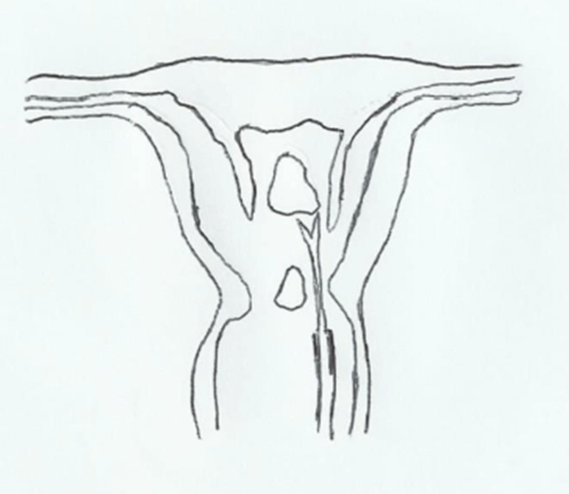
Concomitant laparoscopy is helpful for prevention of uterine perforation but it also cannot visualize the initial point of cutting septal wall (Tajiri et al., 2015). Dye-filled ballooning in a hemiuterus can be easily seen at the opposite site of hemiuterus by hysteroscopy. Pulling the tail of balloon can make the septal wall bulge towards the opposite side. Then we can easily decide which part of septum to cut. Once we make a hole on the septum, we dissect subendometrial area by optic scissors to separate septal endometrium and septal myometrium.
We only cut out septal myometrium by optic scissors leaving septal endometrium intact.
CASE
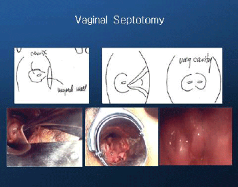
A 40-year-old G1 Po Lo A1 woman was referred to our clinic with 6-year history of infertility. She had 3 cycles of IVF procedures (2 cycles of Controlled Ovarian Stimulation-IVF and 1 cycle of frozen-thawed ET). She had medical history of abortion at early gestation following FET (frozen-thawed-ET). Pelvic exam revealed normal external genitalia, a longitudinal vaginal septum and two cervix (Fig. 1).
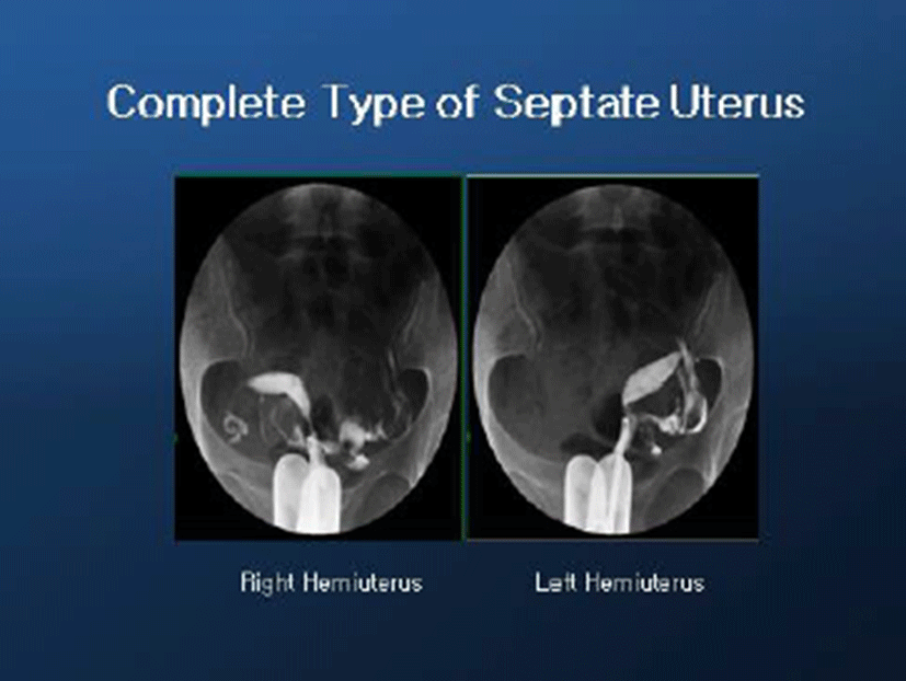
After septotomy of longitudinal vaginal septum, hysterosalpingogram (HSG) was done. It showed two independent hemiuterus and normal spillage of both tubes (Fig. 2).
Transvaginal ultrasonography showed Y shaped uterine cavity, two chambers of upper parts and two fixed lower parts of uterine cavity (Fig. 3).

Before hysteroscopy insertion, we inserted a foley catheter into one hemiuterus and infused indigo calmin (2.0 cc) to make ballooning. Then, we inserted hysteroscope into the other one of hemiuterus so we can cut the bulging area of septal wall. Bulging site of septum was noted and a hole made between two hemiuterus (Figs. 4 and 5).
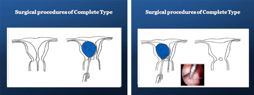
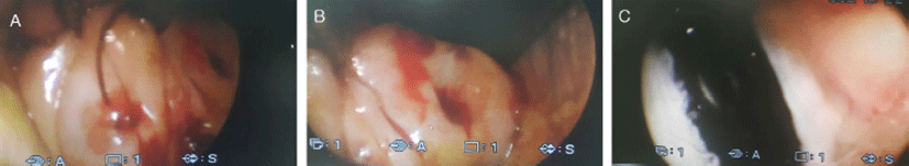
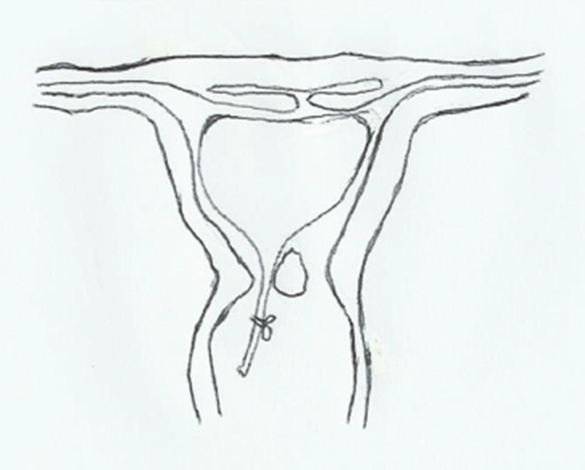
We dissected subendometrial area of septum from the hole and removed septal myometrium and preserved septal endometrium (Fig. 6).
After resection of septal myometrium, we inserted foley balloon catheter and ballooning (3.0 cc) to compress the septal endometrium around the septectomy area (Fig. 7).
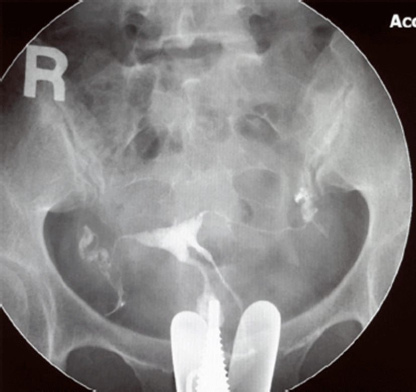
After 1 month, postop HSG was done. HSG shows flat fundal area and two cervical canals (Fig. 8).
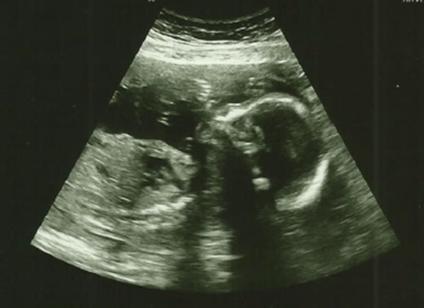
The patient had IVF-ET and FET procedures following the surgery. She had a successful pregnancy and now is at 22 weeks of gestation (Fig. 9).
DISCUSSION
Septate uterus is the most common uterine malformation of uterus. It is due to abnormal resorption of Müllerian canal during embryogenesis (Chan et al., 2011). It is difficult to operate septoplasty in complete type of septate uterus which has thick septal wall and two fixed independent hemiuterus.
When hysteroscopy is inserted into the hemiuterus, it is difficult to evaluate exact septal wall area due to retroversion or anteversion and/or right or left deviation of uterus. Before surgery, MRI is useful to measure length and width of septal wall but it is not good enough to show the initial cutting point of septum. Concomitant laparoscopy during hysteroscopy is also useful to prevent perforation but it cannot visualize the initial point of cutting of septum (Tajiri et al., 2015).
Transabdominal and/or transvaginal ultrasonography may be better in finding the presence of septum is but it is not accurate (Niknejadi et al., 2014). Ballooning in one hemiuterus is helpful in evaluation of septate septal wall. Ballooning makes septum bulge so we can easily evaluate where the septal area is by hysteroscopy. Then we can confirm the septal area by pulling the tail of ballooning. We performed hysteroscopic septoplasty at ambulatory operating room without laparoscopy and without admission.
Laparoscopy may require more manpower, spaces, facilities and surgical instruments. Ambulatory hysteroscopic septoplasty using dye-filled ballooning can be easily done at ambulatory operating room and is a recommendable choice of treatment.