Original research paper

Evaluation of Erythrocyte Morphometric Indices in Juvenile Red Spotted Grouper, Epinephelus akaara under Elevated Water Temperature

Md Mofizur Rahman1,2https://orcid.org/0000-0002-0013-3538, Hea Ja Baek1,https://orcid.org/0000-0002-4578-5919
Author Information & Copyright
1Dept. of Marine Biology, Pukyong National University, Busan 48513, Korea
2Dept. of Fisheries and Marine Science, Noakhali Science and Technology University, Noakhali 3814, Bangladesh
Corresponding Author : Hea Ja Baek, Dept. of Marine Biology, Pukyong National University, Busan 48513, Korea. Tel: +82-51-629-5924, E-mail: hjbaek@pknu.ac.kr

© Copyright 2019 The Korean Society of Developmental Biology. This is an Open-Access article distributed under the terms of the Creative Commons Attribution Non-Commercial License (http://creative-commons.org/licenses/by-nc/3.0/) which permits unrestricted non-commercial use, distribution, and reproduction in any medium, provided the original work is properly cited.

Received: Sep 30, 2019 ; Revised: Oct 12, 2019 ; Accepted: Oct 23, 2019

Published Online: Dec 31, 2019

ABSTRACT

Higher thermal exposure can influence the blood cell morphology and count. Hence, based on the previous results (Rahman et al., 2019), the present study evaluated the morphometric indices of erythrocytes and their nucleus under different water temperatures (25°C, 28°C, 31°C, and 34°C) to investigate their use as an indicator of thermal stress in red spotted grouper, Epinephelus akaara. 180 healthy specimens of E. akaara were exposed to four temperature conditions (25°C as control, 28°C, 31°C, and 34°C) for 42 days, following 2 weeks of acclimation at 25°C. Erythrocyte major axis (EL), erythrocyte minor axis (EW), nucleus major axis (NL), and nucleus minor axis (NW) were examined from the blood smears on each sampling day (i.e., 2, 7, and 42 days of thermal exposure). EL and NL were significantly decreased, whereas EW and NW were increased at higher water temperature (31°C and 34°C). The major-minor axis proportions of erythrocytes and their nucleus (EL/EW; NL/NW) were decreased with increasing water temperature (31°C and 34°C). The strong relationships were observed among the morphometric indices of erythrocytes and their nucleus, especially in EL vs. NL and EW vs. NW. This study reveals that elevated water temperature (31°C and 34°C) can influence the major and minor axis morphometry of erythrocytes and their nucleus in red spotted grouper. These indices may be used as stress indicators to monitor the health status of E. akaara and probably for other fish species.

Keywords: Epinephelus akaara; Temperature; Erythrocyte; Morphometric indices; Stress indicator

INTRODUCTION

Analysis of blood cell can reveal a measurable amount of physiological changes in fish more rapidly than any physiological assessment parameters (Rowan, 2007). It can also respond to changes in other tissues due to exposure of environmental stressors (i.e., temperature, salinity, overcrowding, and water quality) (Ruas et al., 2008). Therefore, examining blood cells has been considered a valuable approach for analyzing the health status of fish and enhancing our understanding of the relationship of blood characteristics with the habitat and adaptability of a species to an environment (Fazio et al., 2012; Shahjahan et al., 2018; De et al., 2019; Islam et al., 2019). Thus, the use of blood indicators in fisheries research is growing rapidly as it is very important in toxicological research, environmental monitoring and predicting of fish health conditions (Barcellos et al., 2004).

Unlike human and most mammals, fish possess an oval-shaped structure of erythrocyte with a condensed nucleus (Jagoe & Welter, 1995). Fish erythrocytes are more responsive to environmental stresses, and often vary in morphology and effectiveness of oxygen transport (Lowe-Jinde & Niimi, 1983). However, variations in erythrocytes and its nuclear size and structure within species are smaller compared to interspecies differences (Najiah et al., 2008; Fang et al., 2014). In our previous study, we examined the blood cell morphology and number of E. akaara under high water temperature exposure and the findings are useful to assess the primary immune response of fish under stressful conditions (i.e., thermal stress) (Rahman et al., 2019).

The red spotted grouper is a sub-tropical species of serranidae family which prefers to inhabit shallower depths of the coastal area (Sadovy & Cornish, 2000). It is a promising aquaculture species due to its high market value in southern Japan, Korea, Hong Kong, Taiwan, and southern China (Morris et al., 2000). The normal range of water temperature in the tropics to which fish are adapted is 25°C–35°C (Howerton, 2001). The optimal water temperature at which E. akaara can live without stress was reported to be 24°C–28°C (Cho et al., 2015; Lee & Baek, 2018). However, temperature can increase to a point that may become harmful for growth and damage physiological processes (Fu et al., 2018). Though a number of studies were conducted to make a better understanding of blood cell morphology of fishes under various environmental toxicants (Rowan, 2007; Osman et al., 2018; Shahjahan et al., 2018; Islam et al., 2019; Rahman et al., 2019), the effect of high temperature on erythrocyte and nuclear morphometry are not well studied. Therefore, the present study was intended to evaluate the morphometric indices of erythrocytes and their nucleus under elevated water temperature which will help to check the health status of E. akaara in a quick time.

MATERIALS AND METHODS

1. Experimental fishes and design

180 healthy and active specimens of E. akaara with an average total length (TL) of 8.28±0.10 cm and body weight (BW) of 8.53±0.27 g were collected from the Marine Science Institute, Jeju National University, Korea. The juveniles were acclimated in aquaria at a water temperature of 25±0.5°C, salinity of 34 psu, dissolved oxygen levels≥6.6 mg/L, pH of 7.8, and a 12L: 12D photoperiod for 2 weeks before the experiment. The fish were fed commercial feed (Otohime Hirame; Marubeni Nisshin Feed Co., Ltd., Tokyo, Japan) twice a day until satiation. The fishes were exposed to four temperature conditions (25°C as control, 28°C, 31°C, and 34°C), each with three replications for 6 weeks. Temperature was gradually increased (Δ1°C/h) using a thermostat (OKE-6422H; OKE, Busan, Korea) to reach the target temperature conditions (25°C, 28°C, 31°C, and 34°C). Approximately 10% of the water was replaced daily with filtered clean seawater as feces and debris were siphoned from the tank.

2. Sampling and erythrocyte morphometry analysis

15 fishes were sampled from each temperature group (i.e., 5 fishes from each aquaria; n=15) on each sampling day (i.e., 2, 7, and 42 days of thermal exposure). Fish were randomly captured and lightly anaesthetized with 300 ppm 2-phenoxyethanol (Sigma Aldrich, St. Louis, MO, USA). Blood was collected from the caudal vein using heparinized capillary tubes, labeled, and stored in 1.5 mL centrifuge tubes. The whole blood was withdrawn less than 1 min per fish to avoid handling stress.

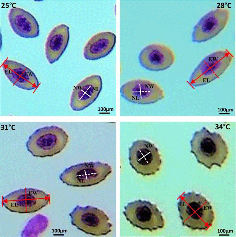
Blood smears were prepared immediately after collection, air-dried for 10 min, fixed in 95% methanol for 5 min, stained with Wright-Giemsa solution, rinsed with distilled water, air-dried and mounted with malinol. Three slides were prepared from each fish and then photos were taken under light microscopy. Erythrocytes in fish are elliptical, and as such, two different diameters are provided: erythrocyte major axis (EL) and erythrocyte minor axis (EW). Nucleus major axis (NL) and nucleus minor axis (NW) were also obtained. On each slide, EL and EW of randomly selected 100 mature erythrocytes and their nuclei (NL and NW) were measured by an Olympus ocular micrometer at a magnification of ×600 (Olympus BX-50, Japan). Erythrocyte and nuclear sizes (ES and NS) were calculated according to formulas [(EL×EW×π)/4] and [(NL×NW×π)/4], respectively (Metin et al., 2008).

3. Statistical analysis

Data have been presented as mean±SEM and analyzed by using SPSS statistics software (ver. 21.0; IBM Corp., Armonk, NY, USA). One-way analysis of variance (ANOVA) followed by Duncan’s multiple range test was used to assess the significant differences among the different temperature groups. Pearson’s correlation coefficient test was conducted to estimate the relationships among the morphometric indices of erythrocyte. In addition, regression analysis and goodness of fit (R2) were determined when there was a significant correlation between tested variables. The results were considered significant at p<0.05.

RESULTS AND DISCUSSION

Elevated water temperature induced significant changes in erythrocytes and nucleus structure of 31°C and 34°C groups, compared to those in the 25°C and 28°C groups. Fishes of 31°C and 34°C groups showed round-shaped erythrocyte structure containing large nucleus, whereas erythrocytes of 25°C and 28°C groups showed oval-shaped structure with condensed nucleus (Fig. 1).

dr-23-4-345-g1
Fig. 1. Measurement of major and minor axis of erythrocyte and nucleus of red spotted grouper, Epinephelus akaraa. Red and white line indicates the diameters of erythrocytes and nucleus, respectively. EL, erythrocyte major axis; EW, erythrocyte minor axis; NL, nucleus major axis; NW, nucleus minor axis. Scale bars=100 μm.
Download Original Figure

The morphometric indices of erythrocytes and their nucleus under different water temperatures (25°C, 28°C, 31°C, and 34°C) are shown in Table 1. Major and minor axis morphometry, and size of erythrocytes and nucleus differed significantly during the thermal exposure period (42 days). The major and minor axis of erythrocytes and nucleus were changed reversely. EL and NL were significantly (p<0.05) decreased, whereas EW and NW were increased at higher water temperature (31°C and 34°C). In addition, the major-minor axis proportion of erythrocytes and their nucleus (EL/EW; NL/NW) were decreased with increasing water temperature (31°C and 34°C). On the other hand, the size of erythrocytes and their nucleus (ES, NS and ES/NS) showed irregular changes under elevated water temperature.

Table 1. Changes in morphometric indices of erythrocytes and nucleus [major and minor axis, and size] in red spotted grouper, Epinephelus akaara exposed to different water temperatures (25°C, 28°C, 31°C, and 34°C) for 42 days
Morphometric indices Temperature groups Period of thermal exposure (days)
2 7 42
EL (μm) 25°C 11.53±0.09a 11.45±0.07a 11.30±0.08a
28°C 11.36±0.02a 11.19±0.07a 10.35±0.22b
31°C 9.62±0.17b 9.02±0.14b 8.68±0.18c
34°C 8.34±0.04c 8.44±0.12c 8.78±0.19c
EW (μm) 25°C 5.59±0.10b 5.59±0.08c 5.77±0.22c
28°C 5.42±0.03b 5.91±0.07b 5.68±0.14c
31°C 6.01±0.02a 6.17±0.09b 6.49±0.07b
34°C 6.03±0.09a 7.16±0.08a 7.36±0.11a
EL/EW 25°C 2.06±0.05a 2.05±0.12a 1.96±0.06a
28°C 2.09±0.09a 1.90±0.08a 1.82±0.05a
31°C 1.60±0.06b 1.47±0.05b 1.34±0.04b
34°C 1.39±0.02c 1.18±0.06c 1.19±0.03c
NL (μm) 25°C 5.12±0.22a 5.48±0.05a 5.12±0.06b
28°C 5.06±0.17a 5.30±0.15a 5.46±0.06a
31°C 4.66±0.21a 4.15±0.04b 4.47±0.06c
34°C 3.55±0.14b 4.21±0.19b 4.49±0.10c
NW (μm) 25°C 2.94±0.07b 3.32±0.08c 2.65±0.06c
28°C 3.05±0.10b 3.20±0.03c 3.28±0.12b
31°C 3.51±0.08a 3.83±0.02b 3.68±0.24b
34°C 3.73±0.08a 4.00±0.03a 4.28±0.09a
NL/NW 25°C 1.74±0.09a 1.65±0.03a 1.94±0.07a
28°C 1.67±0.02a 1.66±0.04a 1.71±0.02b
31°C 1.33±0.04b 1.09±0.06b 1.20±0.04c
34°C 0.95±0.05c 1.05±0.04b 1.05±0.04d
ES (μm2) 25°C 50.65±0.65a 50.18±0.62b 51.28±0.88a
28°C 48.39±2.46b 51.98±0.28a 46.24±0.88b
31°C 45.55±3.88ab 43.98±0.48d 44.25±0.53b
34°C 39.44±0.50b 47.44±0.14c 50.78±1.53a
NS (μm2) 25°C 11.83±0.54a 14.27±0.12a 10.65±0.13c
28°C 12.14±0.23a 13.27±0.04b 13.83±0.33b
31°C 12.95±0.39a 12.49±0.57b 13.19±0.26b
34°C 10.43±0.12b 13.22±0.15b 15.13±0.24a
ES/NS 25°C 4.30±0.22a 3.53±0.09b 4.82±0.21a
28°C 4.02±0.26ab 3.91±0.08a 3.44±0.28b
31°C 3.55±0.20b 3.50±0.04b 3.41±0.06b
34°C 3.81±0.14ab 3.59±0.11b 3.41±0.21b

Sampling was done at three different time points (2, 7, and 42 days). Values are mean±SEM of three replicates (n=15; 5 fish per tank). 100 erythrocytes and their nucleus were measured from each blood smeared slide.

Different lowercase letters indicate the significant difference between groups at equivalent times (ANOVA, Duncan’s multiple range test; p<0.05).

EL, erythrocyte major axis; EW, erythrocyte minor axis; NL, nucleus major axis; NW, nucleus minor axis; ES, erythrocyte size; NS, nucleus size.

Download Excel Table

Both positive and negative correlations were observed among the morphometric indices of erythrocytes and nucleus of red spotted grouper (Table 2 and Fig. 2a–f). A highly significant positive relationship was found in EL vs. NL and EW vs. NW, whereas significant negative relationship was detected in EL vs. EW, NL vs. NW, EL vs. NW, and EW vs. NL.

dr-23-4-345-g2
Fig. 2. Linear correlations among the morphometric indices of erythrocytes and their nucleus in red spotted grouper, Epinephelus akaraa. (a) Erythrocyte major axis (EL) vs. Erythrocyte minor axis (EW); (b) Nucleus major axis (NL) vs. Nucleus minor axis (NW); (c) Erythrocyte major axis (EL) vs. Nucleus major axis (NL); (d) Erythrocyte minor axis (EW) vs. Nucleus minor axis (NW); (e) Erythrocyte major axis (EL) vs. Nucleus minor axis (NW); (f) Erythrocyte minor axis (EW) vs. Nucleus major axis (NL).
Download Original Figure
Table 2. Pearson correlation coefficient and the regression equation for the morphometric indices of erythrocytes and nucleus in red spotted grouper, Epinephelus akaara
Paired samples Correlation coefficient (r)
EL (x) vs. EW (y) −0.740**
NL (x) vs. NW (y) −0.617**
EL (x) vs. NL (y) 0.830**
EW (x) vs. NW (y) 0.796**
EL (x) vs. NW (y) −0.814**
EW (x) vs. NL (y) −0.536**

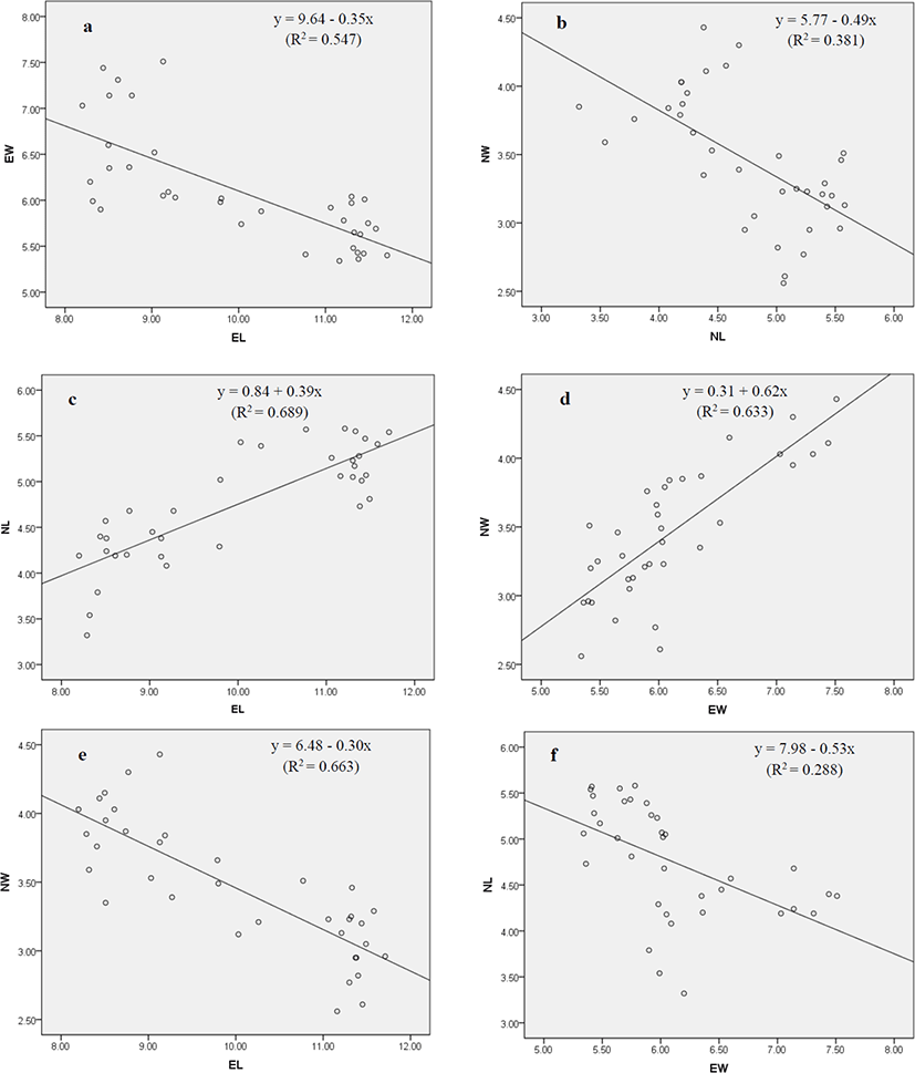
See the regression plot (Fig. 2a-f) for each paired samples **p<0.01.

EL, erythrocyte major axis; EW, erythrocyte minor axis; NL, nucleus major axis; NW, nucleus minor axis; ES, erythrocyte size; NS, nucleus size.

Download Excel Table

Since erythrocytes are influenced by a variety of environmental stressors, they have the potential to be used as stress indicators. Their evaluation in fish has become an important means of understanding the toxicological impacts of exposure hazards (Borges et al., 2007). In this study, morphometric indices of erythrocytes and their nucleus were evaluated under different water temperatures (25°C, 28°C, 31°C, and 34°C) to investigate their use as an indicator of thermal stress in red spotted grouper.

In the present study, changes in erythrocytes and their nucleus structure were observed in E. akaara under elevated water temperature (31°C and 34°C). In our previous study, we reported that thermal stress causes various types of erythrocytic cellular abnormalities (ECA) in E. akaara (Rahman et al., 2019). The erythrocytes of 25°C and 28°C were found in oval to elliptical shape, but turned into round shape when exposed to 31°C and 34°C. These altered structure in erythrocytes and nucleus can induce anemic condition to the thermally stressed fishes. Similar abnormalities in erythrocyte and nuclei were reported in striped catfish, Pangasianodon hypophthalmus when treated with higher temperature (36°C) for 7–28 days (Shahjahan et al., 2018; Islam et al., 2019). The observed structural changes in erythrocyte and nuclei may be attributed to one or more of the following factors: (i) unequal distribution of hemoglobin (Mekkawy et al., 2011); (ii) necrosis of the red blood cells (Ateeq et al., 2002); (iii) interruption in lipid solubility to erythrocyte’s membrane (Walia et al., 2013).

Fish erythrocytes and their nucleus have a wide range of shape and sizes among different species (Jagoe & Welter, 1995). The changes in erythrocytes depend on the fish species, age, health and the environment where the fish live (Vazquez & Guerrero, 2007; Zexia et al., 2007; Motlagh et al., 2012). Measurement of morphometric indices of erythrocytes and nucleus [major axis, minor axis, and size] under different water temperatures (25°C, 28°C, 31°C, and 34°C) demonstrated that erythrocytes and their nucleus varied in size at higher temperature (31°C and 34°C) which is the clear indication of stress. The observed decrease in major axis (EL, NL) and increase in minor axis (EW, NW) of erythrocytes and their nucleus might be because of changing the erythrocyte’s shape from elongate to round which causes the reduction in major axis and enlargement in minor axis. The observed variation could also be explained by the following factors: (i) differing the dissolved oxygen levels in experimental groups; (ii) the maturation stage of the erythrocytes; (iii) the sample size of research. These findings are consistent with those of Rowan (2007), who reported that erythrocyte and nucleus morphometry of brown bullheads (Ameiurus nebulosus) were affected due to the pollution of river water by agricultural runoffs or industrial effluents. Similar results were also reported in Nile tilapia (Oreochromis niloticus niloticus) and African catfish (Clarias gariepinus) collected from contaminated sites of the river Nile (Osman et al., 2018). In this study, the strong relationships were observed among the morphometric indices of erythrocytes and their nucleus. Hardie & Hebert (2003) reported that erythrocytes and nuclear sizes are significantly correlated in ray-finned fishes, cartilaginous fishes and all species combined.

In conclusion, the results of the present study reveal that elevated water temperature (31°C and 34°C) beyond the optimal range (24°C–28°C) affects the morphometric indices of erythrocytes and their nucleus, especially EL, EW, NL, and NW which can be considered as stress indicators (i.e., thermal stress) of red spotted grouper. This fine information may be helpful to monitor the health status of E. akaara and probably for other fish species.

CONFLICT OF INTEREST

The authors declare no potential conflict of interest.

ACKNOWLEDGEMENTS

This research was supported by a grant (213008-05-3-WT511) from the Korean Ministry of Agriculture, Food and Rural Affairs (MAFRA), the Korean Ministry of Oceans and Fisheries (MOF), the Korean Rural Development Administration (RDA), and the Korea Forest Service (KFS).

AUTHOR CONTRIBUTIONS

Conceptualization: Baek HJ.

Data curation: Rahman MM.

Formal analysis: Rahman MM.

Methodology: Rahman MM, Baek HJ.

Software: Rahman MM.

Validation: Rahman MM, Baek HJ.

Investigation: Rahman Mm.

Writing - original draft: Rahman MM, Baek HJ.

Writing - review & editing: Rahman MM, Baek HJ.

ETHICS APPROVAL

This article does not require IRB/IACUC approval because there are no human and animal participants.

REFERENCES

1.

Ateeq B, Abulfarah M, Ali MN, Ahmad W. 2002; Induction of micronuclei and erythrocyte alterations in the catfish Clarias batrachus by 2,4-dichlorophenoxyacetic acid and butachlor. Mutat Res. 518:135-144

2.

Barcellos LJG, Kreutz LC, de Souza C, Rodriguez LB, Fioreze I, Quevedo RM, Cericato L, Soso AB, Fagundes M, Conrad J, Lacerda LA, Terra S. 2004; Hematological changes in Jundia (Rhamdia quelen) after acute and chronic stress caused by usual aquacultural management, with emphasis on immunosuppressive effects. Aquaculture. 237:229-236

3.

Borges A, Scotti LV, Siqueira DR, Zanini R, Do Amaral F, Jurinitz DF, Wassermann GF. 2007; Changes in hematological and serum biochemical values in Jundia Rhamdia quelen due to sub-lethal toxicity of cypermethrin. Chemosphere. 69:920-926

4.

Cho HC, Kim JE, Kim HB, Baek HJ. 2015; Effects of water temperature change on the hematological responses and plasma cortisol levels in growing of red spotted grouper, Epinephelus akaara. Dev Reprod. 19:19-24

5.

De M, Ghaffar MA, Noor NM, Cob ZC, Bakar Y, Das SK. 2019; Effects of water temperature and diet on blood parameters and stress levels in hybrid grouper (Epinephelusfuscoguttatus♀× Elanceolatus♂) juveniles. Aquac Rep. 15:100219

6.

Fang J, Chen K, Cui HM, Peng X, Li T, Zuo ZC. 2014; Morphological and cytochemical studies of peripheral blood cells of Schizothorax prenanti. Anat Histol Embryol. 43:386-394

7.

Fazio F, Filiciotto F, Marafioti S, Di Stefano V, Assenza A, Placenti F, Buscaino G, Piccione G, Mazzola S. 2012; Automatic analysis to assess haematological parameters in farmed gilthead sea bream (Sparus aurata Linnaeus, 1758). Mar Freshw Behav Physiol. 45:63-73

8.

Fu KK, Fu C, Qin YL, Bai Y, Fu SJ. 2018; The thermal acclimation rate varied among physiological functions and temperature regimes in a common cyprinid fish. Aquaculture. 495:393-401

9.

Hardie DC, Hebert PDN. 2003; The nucleotypic effects of cellular DNA content in cartilaginous and ray-finned fishes. Genome. 46:683-706

10.

Howerton R. 2001 Best management practices for Hawaiian aquaculture. Center for Tropical and Subtropical Aquaculture, Waimanalo, Publication No. 148. .

11.

Islam MA, Uddin MH, Uddin MJ, Shahjahan M. 2019; Temperature changes influenced the growth performance and physiological functions of Thai pangas Pangasianodon hypophthalmus. Aquac Rep. 13:100179

12.

Jagoe CH, Welter DA. 1995; Quantitative comparisons of the morphology and ultrastructure of erythrocyte nuclei from seven freshwater fish species. Can J Zool. 73:1951-1959

13.

Lee JW, Baek HJ. 2018; Determination of optimal temperature(s) in juvenile red‐spotted grouper Epinephelus akaara (Temminck & Schlegel) based on growth performance and stress responses. Aquac Res. 49:3228-3233

14.

Lowe-Jinde L, Niimi AJ. 1983; Influence of sampling on the interpretation of haematologicalmeasurements of rainbow trout, Salmo gairdneri. Can J Zool. 61:396-402

15.

Mekkawy IA, Mahmoud UM, Sayed AEH. 2011; Effects of 4-nonylphenol on blood cells of the African catfish Clarias gariepinus (Burchell, 1822). Tissue Cell. 43:223-229

16.

Metin K, Koca YB, Kiral FK, Koca S, Turkozan O. 2008; Blood cell morphology and plasma biochemistry of captive Mauremys caspica (Gmelin, 1774) and Mauremys rivulata (Valenciennes, 1833). Acta Vet Brno. 77:163-174

17.

Morris AV, Roberts CM, Hawkins JP. 2000; The threatened status of groupers (Epinephelinae). Biodivers Conserv. 9:919-942

18.

Motlagh SP, Zarejabad AM, Nasrabadi RG, Ahmadifar E, Molaee M. 2012; Haematology, morphology and blood cells characteristics of male and female Siamese fighting fish (Bettasplendens). Comp Clin Pathol. 21:15-21

19.

Najiah M, Nadirah M, Marina H, Lee SW, Nazaha WH. 2008; Quantitative comparisons of erythrocyte morphology in healthy freshwater fish species from Malaysia. Res J Fish Hydrobiol. 3:32-35.

20.

Osman AGM, AbouelFadl KY, Abd El Reheem AEM, Mahmoud UM, Kloas W, Moustafa MA. 2018; Blood biomarkers in Nile tilapia Oreochromis niloticus niloticus and African catfish Clarias gariepinus to evaluate water quality of the river Nile. J Fish Sci Com. 12:1-15

21.

Rahman MM, Kim HB, Baek HJ. 2019; Changes in blood cell morphology and number of red spotted grouper, Epinephelus akaara in response to thermal stress. Dev Reprod. 23:139-148

22.

Rowan MW. 2007 Use of blood parameters as biomarkers in brown bullheads (Ameiurus nebulosus) from Lake Erie tributaries and Cape Cod ponds. Doctoral dissertation. The Ohio State University. .

23.

Ruas CBG, Carvalho CD, de Araujo HSS, Espindola ELG, Fernandes MN. 2008; Oxidative stress biomarkers of exposure in the blood of cichlid species from a metal-contaminated river. Ecotox Environ Safe. 71:86-93

24.

Sadovy Y, Cornish AS. 2000 Reef fishes of Hong Kong. Hong Kong University Press. Hong Kong: .

25.

Shahjahan M, Uddin MH, Bain V, Haque MM. 2018; Increased water temperature altered hemato-biochemical parameters and structure of peripheral erythrocytes in striped catfish Pangasianodon hypophthalmus. Fish Physiol Biochem. 44:1309-1318

26.

Vazquez GR, Guerrero GA. 2007; Characterization of blood cells and hematological parameters in Cichlasoma dimerus (Teleostei, Perciformes). Tissue Cell. 39:151-160

27.

Walia GK, Handa D, Kaur H, Kalotra R. 2013; Erythrocyte abnormalities in a freshwater fish, Labeo rohita exposed to tannery industry effluent. Int J Pharm Biol Sci. 3:287-295.

28.

Zexia G, Weimin W, Yi Y, Abbas K, Dapeng L, Guiwei Z, Diana JS. 2007; Morphological studies of peripheral blood cells of the Chinese sturgeon, Acipenser sinensis. Fish Physiol Biochem. 33:213-222